

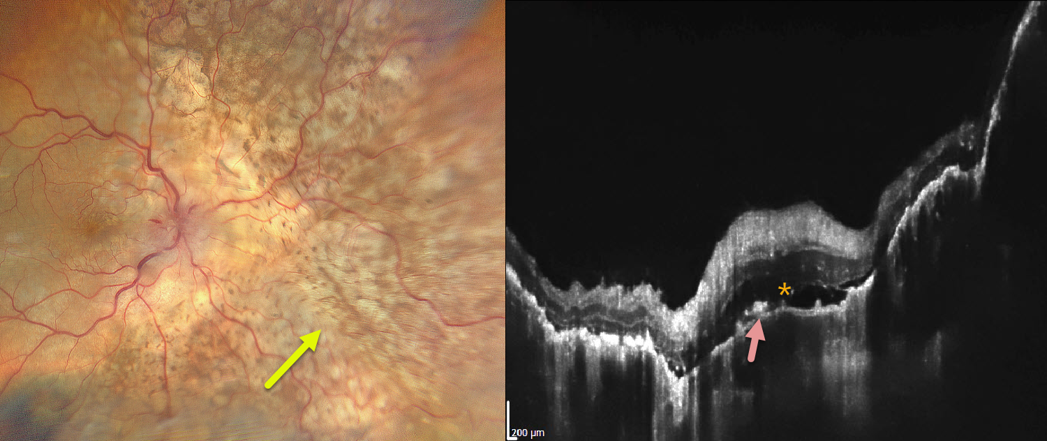
This 68YO male presented with severe vision loss in his right eye for several weeks. There was a known history of metastatic lung adenocarcinoma. Vision was light perception OD and 20/20 in his normal OS.
Color photography shows variably confluent, creamy-yellow subretinal lesions scattered within the macula and extending into the midperiphery. OCT scanning shows an undulating thickened choroid with shallow overlying subretinal fluid and scattered hyperreflective material above the RPE, likely representing lipofuscin.
Learning Points:
Although uveal melanoma is the most common primary intraocular malignancy, metastatic choroidal tumors are the most common intraocular malignancies. Lung and breast cancer are the most common sources.



