

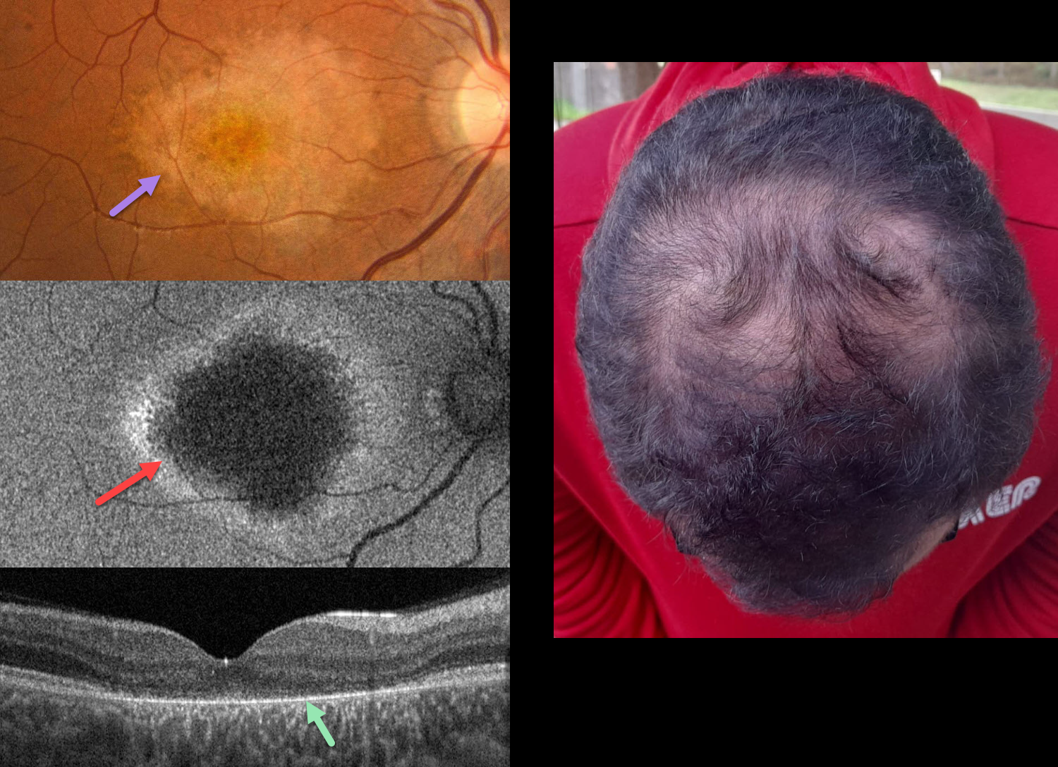
This 19YO male was referred for progressive vision loss and hemeralopia since early childhood. Ocular family history was negative, although his parents were consanguineous. Vision was 20/40 OU.
Color photography of his right eye shows an oval to tear-drop shaped area of central and nasal macular atrophy with clumps of more distal subretinal pigmentation. Fundus autofluorescence (FAF) shows central hypo-FAF with a ring of peripheral hyper-FAF. OCT scanning shows near-complete loss of the outer retinal bands external to the outer nuclear layer. Similar findings were noted in his left eye (not shown). The hair is thinned along the top of his head. Genetic testing revealed a likely pathogenic homozygous variant in the CDH3 gene (16q22.1).
Learning Points:
Hypotrichosis with juvenile macular dystrophy is a rare autosomal recessive disorder caused by biallelic mutations in the CDH3 gene, which encodes for P-cadherin, a calcium-dependent cell adhesion protein expressed in hair follicles and RPE. It is characterized by congenital sparse, short scalp hair (with normal facial and body hair). Progressive macular chorioretinal atrophy begins in childhood, often causing severe central visual impairment by the second to third decade of life.
This case was previously published in Ophthalmology Retina 2021;5:593.



