

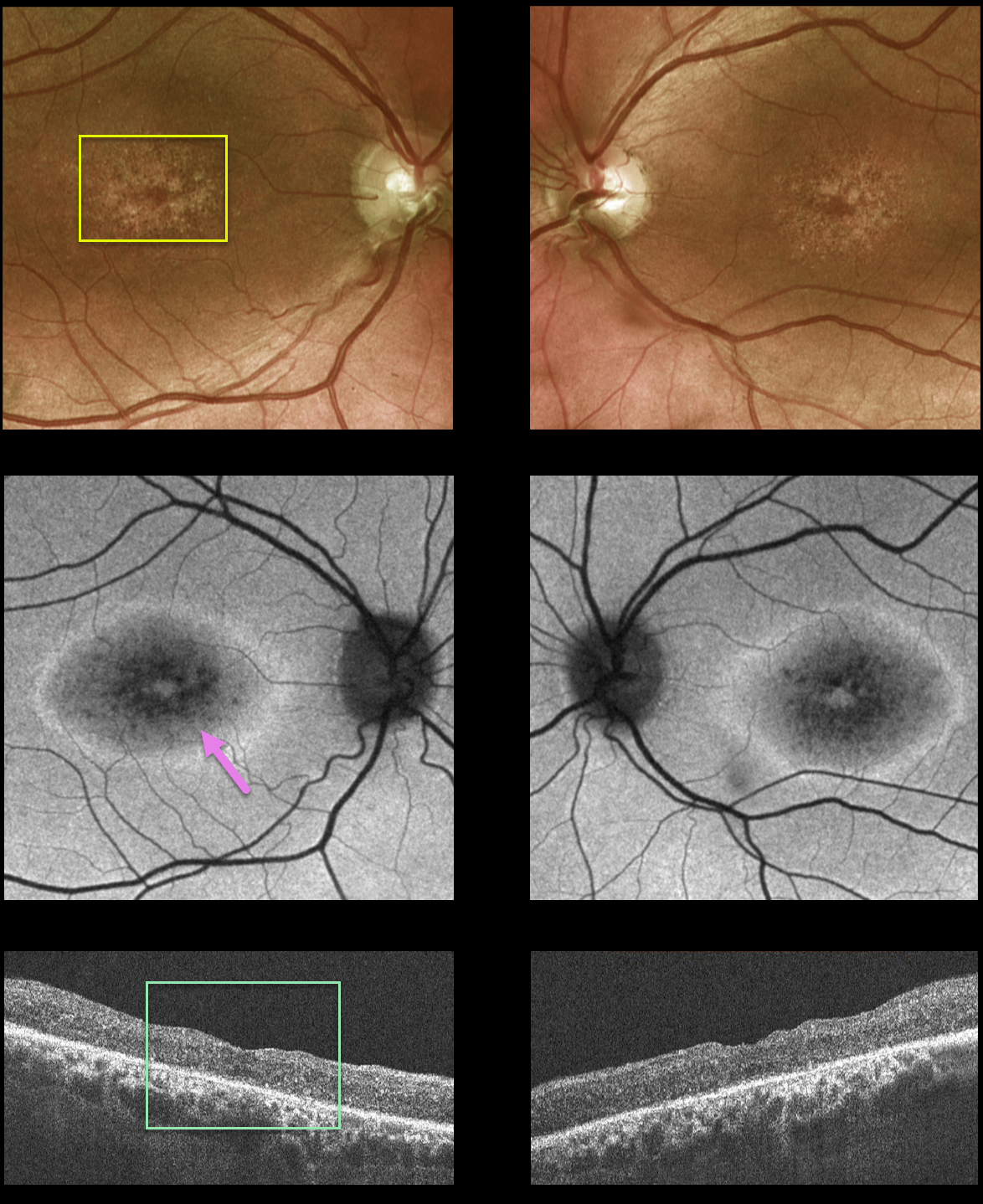
This 42YO male presented to our clinic with a history of X-linked retinoschisis diagnosed elsewhere. Vision was 20/70 OD and 20/200 OS.
Fundus photos show atrophic foveal pigmentary changes. Fundus autofluorescence (FAF) shows mottled central hypo-FAF with a ring of surrounding hyper-FAF. OCT scanning shows bilateral macular thinning with disorganization of the normal macular hyper- and hyporeflective bands.
Genetic testing confirmed the presence of a pathogenic RS1 mutation, which is associated with sex-linked retinoschisis.
Learning Points:
Although historically taught to involve the nerve fiber layer, sex-linked retinoschisis in the macula causes schisis throughout the macula, including the inner nuclear, outer nuclear, and outer plexiform layers (see Yu et al, AJO 2010;149:973-978).
Over time, the schisis often collapses with secondary atrophy, as occurred in our patient (see Menke et al, Retina 2011;31:1186-1192).



