

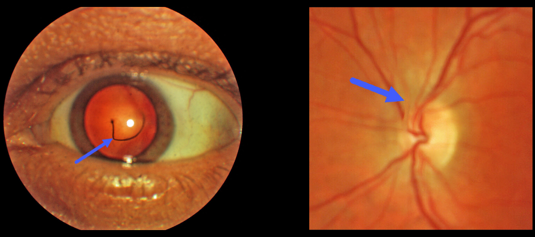
This 44YO male was referred for an asymptomatic vitreous floater. Upon examination, a large translucent strand was noted posterior to the lens, connecting the posterior capsule of the lens to the optic nerve.
Learning Points:
The hyaloid vascular system, embryologically located within the hyaloid canal, provides the developing anterior segment with nutrients. The hyaloid artery usually regresses around 10 weeks of gestation, but in some cases, it may not fully regress.
An anterior remnant may remain on the posterior capsule, which is known as a Mittendorf Dot. Posterior remnants, attached to the optic disc, are otherwise known as Bergmeister’s papilla.



