

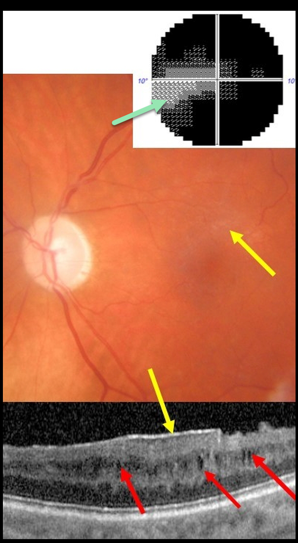
This 61YO male presented with end-stage glaucoma OS, 20/30 vision, and an IOP of 8 mmHg following successful trabeculectomy ten years earlier. He had prior cataract surgery in this eye 15 years ago with no documented history of pseudophakic cystoid macular edema.
There was a moderate macular pucker noted clinically and on OCT, along with multiple inner nuclear layer (INL) microcysts on OCT scanning. He has a tiny island remaining in his visual field.
Learning Points:
Microcystic macular changes have been associated with both advanced primary open-angle glaucoma and idiopathic macular pucker (see Govetto et al, AJO 2017;181:156-165).
Macular pucker-induced mechanical stress, coupled with retrograde trans-synaptic degeneration of bipolar cells secondary to retinal ganglion cell loss, likely predisposes towards the accumulation of fluid within the inner retinas of these patients.
Relative to non-glaucomatous eyes, microcystic macular changes in glaucomatous eyes tend to be more persistent following membrane peeling. This is likely due to retrograde trans-synaptic degeneration.
It is important that these microcystic spaces in eyes with optic neuropathy are not mistaken for inflammatory edema following membrane peeling, which could lead to unnecessary treatment.
Finally, disproportionate preservation of overall retinal thickness despite severe thinning of the ganglion cell and/or nerve fiber layers should not be surprising in glaucomatous eyes.



