

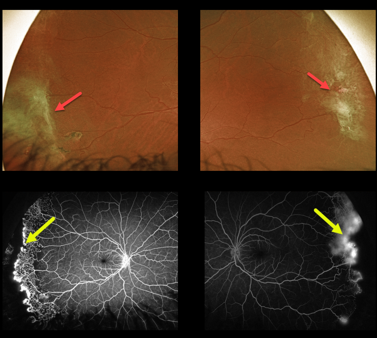
This 43YO female with a known history of SC disease presented with these asymptomatic fundus changes captured with Optos ultra-widefield imaging. Fibrosed areas of peripheral neovascularization are noted in each temporal periphery.
Fluorescein angiography shows a broad, well-demarcated zone of temporal peripheral retinal neovascularization at the junction of perfused and nonperfused retina.
Prophylactic scatter laser to the areas of capillary loss was recommended.
Learning Points:
Patients with sickle cell disease develop symptoms throughout their bodies as stiff, sickled red blood cells block capillary beds.
In the eye, this can cause proliferative sickle cell retinopathy (PSR), with sea-fan-shaped areas of peripheral retinal neovascularization developing at the junction of perfused and ischemic retina. Complications include vitreous hemorrhage and traction retinal detachment.
Although patients with SS disease tend to have more systemic complications, SC disease is usually associated with more severe ocular findings.
Treatment with peripheral scatter laser photocoagulation is somewhat controversial because, unlike more typical proliferative retinopathies (i.e., those from diabetes and branch retinal vein occlusions), these new vessels tend to auto-infarct.
For a great review article on sickle cell disease and the eye, see Elagouz et al, Surv Ophthalmology 2010;55:359-377.



