

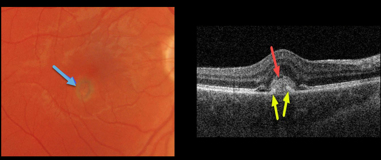
This 30YO male presented with acute unilateral vision loss of 20/50 in his right eye. Ocular examinations were normal bilaterally except for a small area of retinal edema with a subretinal pigment ring in the right inferotemporal fovea.
Swept-source OCT shows a hyperreflective macular neovascularization (MNV) mostly above the RPE. Elevated RPE partially extends into the midportion of the MNV.
In this case, due to our patient’s acute symptoms, we chose to begin anti-VEGF therapy.
Learning Points:
Type 2 MNV often have RPE cells that migrate along its anterior surface, and this appears clinically as a pigment ring. Although the RPE never completely envelops the MNV, its presence often helps to stabilize the lesion’s exudation. In this case, due to our patient’s acute symptoms, we chose to begin anti-VEGF therapy.



