

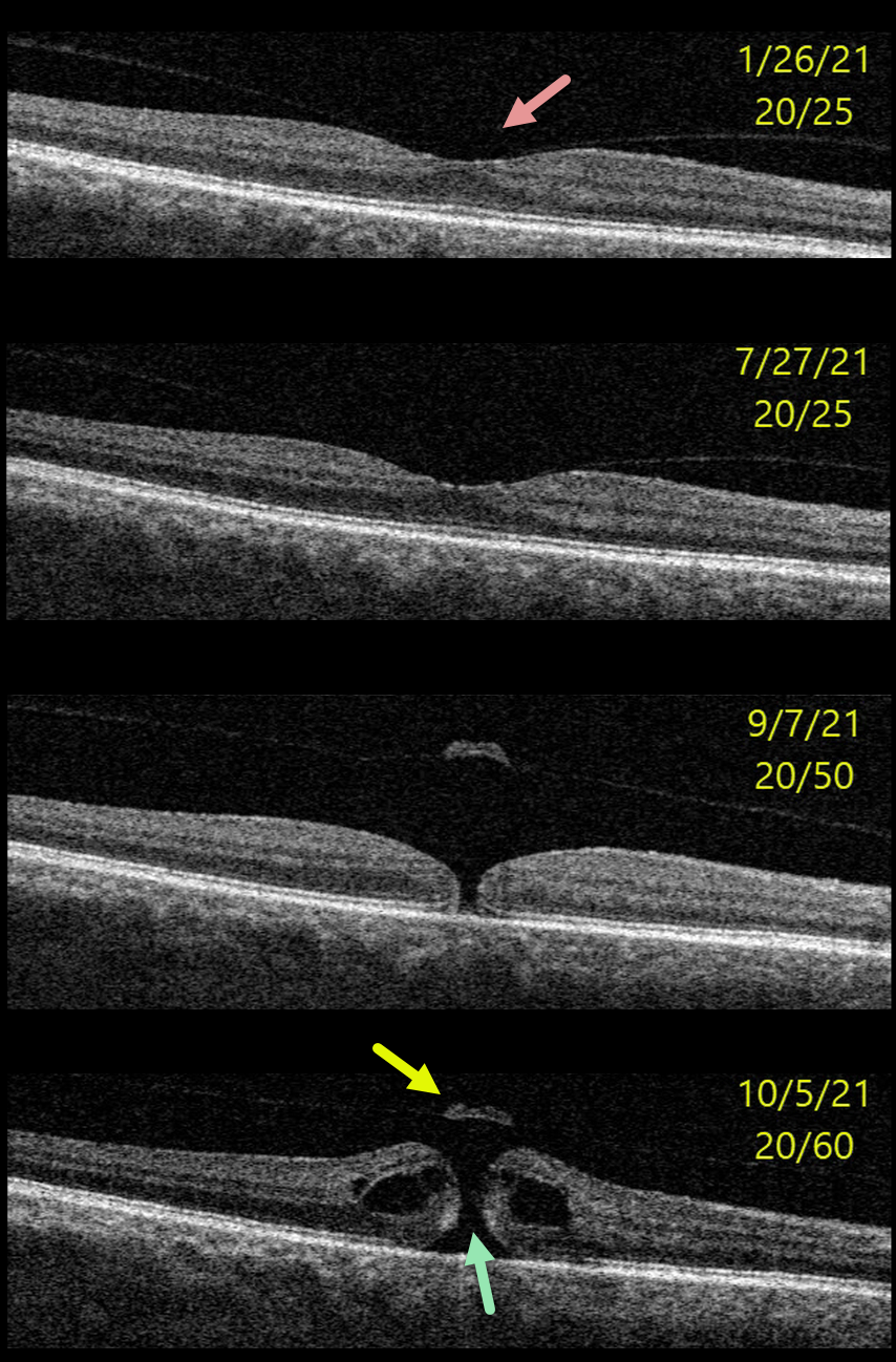
This 64YO male presented with 20/25 vision and asymptomatic vitreomacular adhesion (VMA) in his right eye.
Over the next 8 months, this progressed to subtle focal vitreomacular traction (VMT) and eventually a small full-thickness macular hole without traction. Vitrectomy surgery was recommended.
Learning Points:
In 1988, Dr. Gass described his concept for the pathogenesis of idiopathic macular holes (Arch Ophthalmol 1988;106:629-639).
His classification system, from stage 1 for an impending macular hole through stage 4 for a fully developed macular hole with complete posterior vitreous detachment, was based on his concept of localized, mostly tangential traction caused by shrinking foveal vitreous cortex. This classic paper heralded the advent of modern macular hole surgery for a previously untreatable condition.
The advent of OCT technology, however, showed that macular hole development is driven more by focal vitreomacular traction, as our case beautifully illustrates.
The current classification system, described by the International Vitreomacular Traction Study Group, emphasizes the various characteristics that determine choice of treatment and visual prognosis (Ophthalmology 2013;120:2611-2619). This includes size (small <= 250 microns, medium >250 microns and <= 400 microns, large >400 microns), presence or absence of VMT, and cause (primary from VMT or secondary).



