

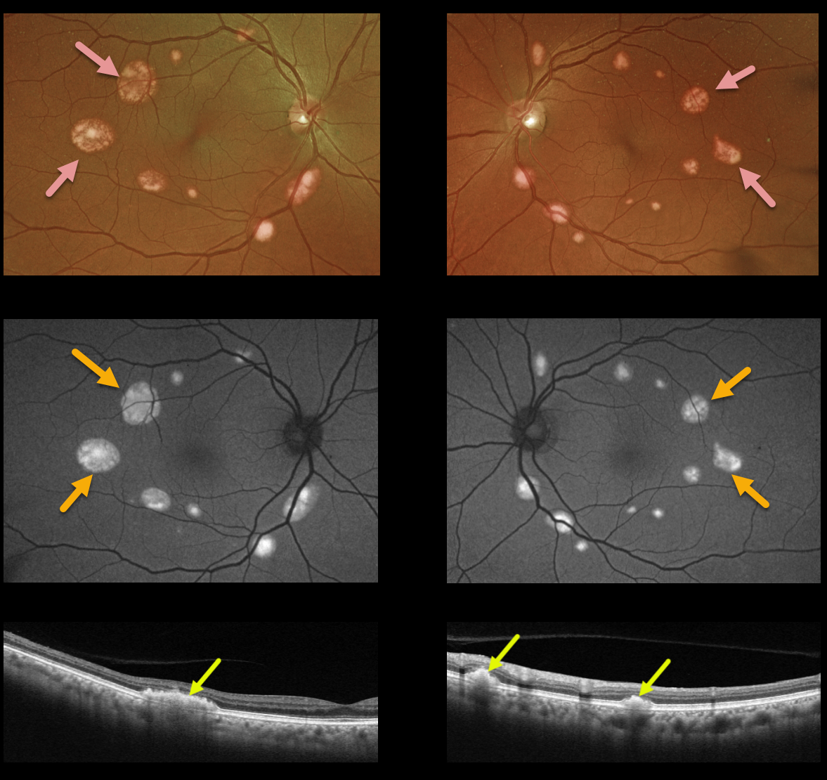
This 68YO female presented with these asymptomatic multifocal subretinal yellow posterior pole lesions. Vision is 20/20 OU.
The sharply demarcated yellowish lesions are markedly hyper-autofluorescent and are hyperreflective on Triton swept-source OCT. There was no known family history, but genetic testing revealed a heterozygous pathogenic BEST1 mutation.
Learning Points:
Best disease is associated with a mutation in the BEST1 gene, which encodes the bestrophin-1 protein. Bestrophin-1, a calcium-activated chloride channel, is primarily found in the basolateral plasma membrane of the RPE.
BEST1 mutations cause a variety of phenotypes, including autosomal recessive bestrophinopathy, best vitelliform macular dystrophy, and autosomal dominant vitreoretinochoroidopathy. Multifocal lesions are a rare phenotype and are not usually associated with subfoveal lesions.
Multifocal Best needs to be differentiated from other causes of multifocal serous detachments, including autosomal recessive bestrophinopathy (see Boon et al, Ophthalmology 2013;120:809-820) and idiopathic or paraneoplastic acute exudative polymorphous vitelliform maculopathy (see Barbazetto et al, Ophthalmology 2018;125:75-88).



