

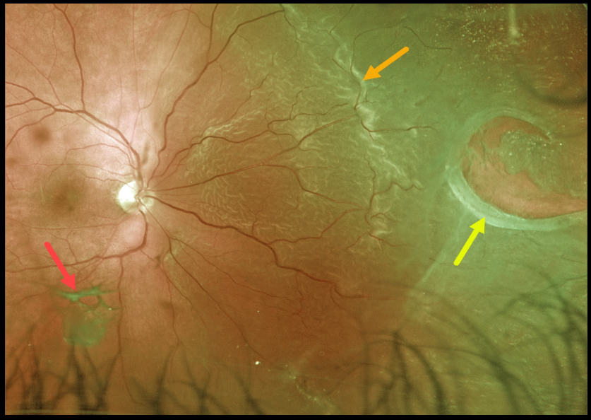
This patient presented with a macula-on rhegmatogenous retinal detachment (RRD) from a large nasal retinal tear. The tear has a rolled posterior edge, indicating possible early proliferative vitreoretinopathy.
The hydration outer retinal folds at the posterior edge of the detachment are one of the key clinical findings that help differentiate a rhegmatogenous from exudative and traction retinal detachments.
The causative posterior vitreous detachment is seen floating above the retina just inferior to the macula.
Learning Points:
The normal neurosensory retina remains attached to the RPE due to numerous physiological mechanisms, including the inner (retinal vascular endothelium) and outer (tight junctions at the apex of the RPE cells) blood retinal barriers, hyperosmotic choroid passively drawing fluid into the subretinal space, RPE pump, interphotoreceptor matrix, and friction between the RPE microvilli and the photoreceptor outer segments.
Rhegmatogenous retinal detachment (RRD) occurs when liquefied vitreous, entering the subretinal space through a retinal break, overwhelms the forces that favor retinal detachment.
Repairing RRD involves finding and closing all breaks, essentially denying the liquid vitreous access to the subretinal space. The break(s) can be closed internally with a gas bubble with pneumatic retinopexy or vitrectomy surgery, or externally by sewing a piece of silicone material to the eye wall (scleral buckle). This indents, or buckles, the sclera up against the retina break.



