

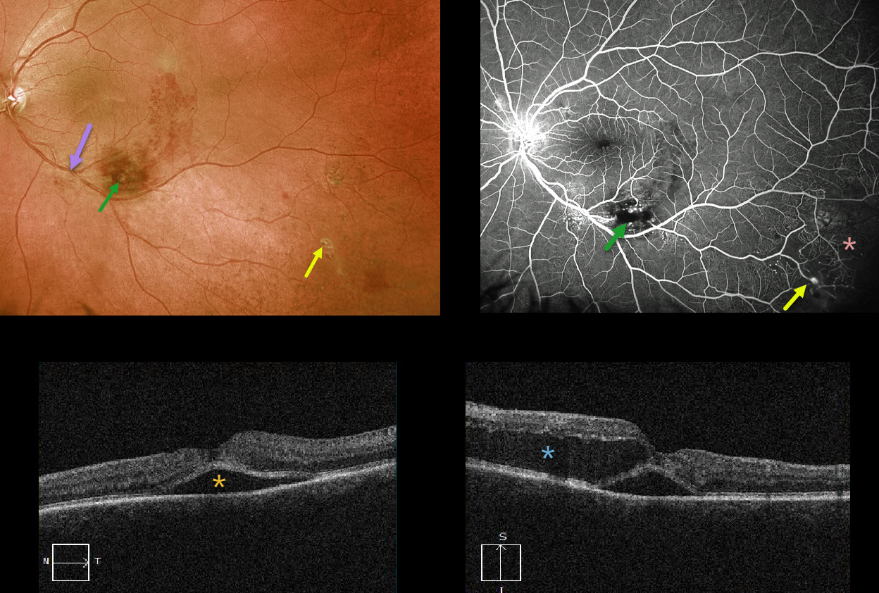
This 59YO female presented with acute vision loss of 20-100 in her left eye. At first glance, it looks like a macular branch retinal vein occlusion (BRVO) arising from an artery crossing over a vein more proximally. There is a likely macroaneurysm within the area of more confluent inferior macular blood.
But a more careful inspection shows some retinal telangiectasia more inferiorly and proximally, as well as more distal inferotemporal areas of retinal hemorrhages, telangiectasia, and an arterial macroaneurysm.
Fluorescein angiography better shows these vascular changes and broad peripheral areas of inferotemporal capillary loss.
OCT shows foveal subretinal fluid with a large pocket of outer intraretinal fluid.
Our best guess is that this patient has Coats disease, given the multiple macroaneurysms, multifocal areas of retinal vascular telangiectasia, and capillary loss. Although uncommon, Coats can be found in females.



