

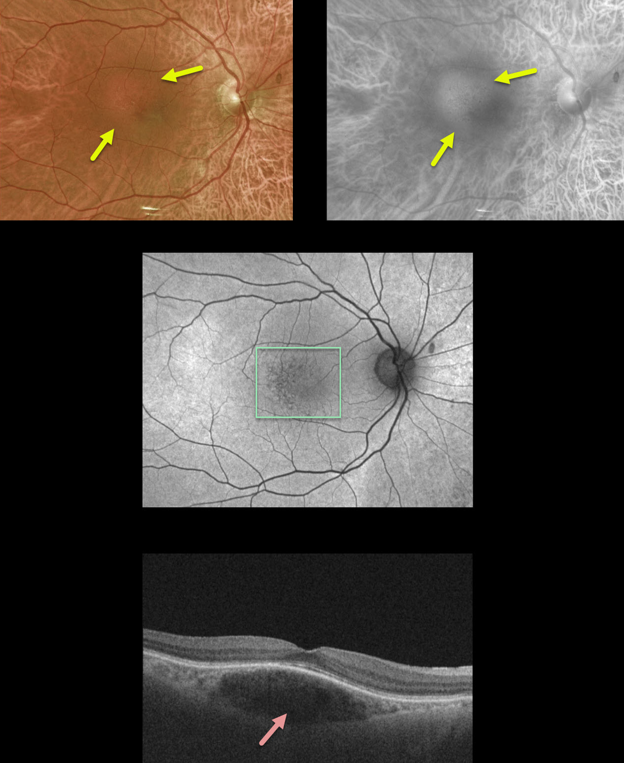
This 44yo female presented with 20/20 vision and no visual complaints.
If you look closely at the color Optos image, you can see a small 1.5 disc diameter subretinal lesion in the superotemporal macula with mild overlying pigmentary changes. The Optos red channel best illuminates the lesion, indicating its choroidal location.
Fundus autofluorescence shows very mild overlying pigmentary changes. OCT shows a somewhat fusiform-shaped hyporeflective choroidal tumor without overlying exudation.
Since our patient was asymptomatic without fluid, we are following her twice yearly and will consider treatment if she develops symptomatic fluid.
Learning Points:
Isolated choroidal hemangiomas are benign vascular lesions. Although they have no malignant potential, they can cause vision loss due to exudation and subretinal and intraretinal fluid, which can be treated with photodynamic therapy.



