

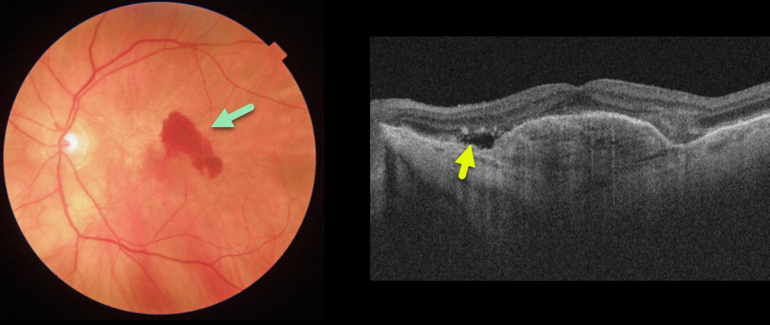
This patient with wet age-related macular degeneration (AMD) was being treated with PRN anti-VEGF injections. She came in for her regular examination without new symptoms and with 20/70 vision.
Although the OCT line scan shows some fairly subtle new sub-retinal fluid, the color photo dramatically shows new blood.
Learning Points:
There is currently some debate about the utility of performing full dilated examinations when patients are being followed with ongoing anti-VEGF therapy.
New blood is quite easy to miss on an OCT without a fundus photograph or examination. In our hands, having a high-quality fundus photograph at each visit, regardless of whether the patient is in for just an injection or a full examination, is invaluable.



