

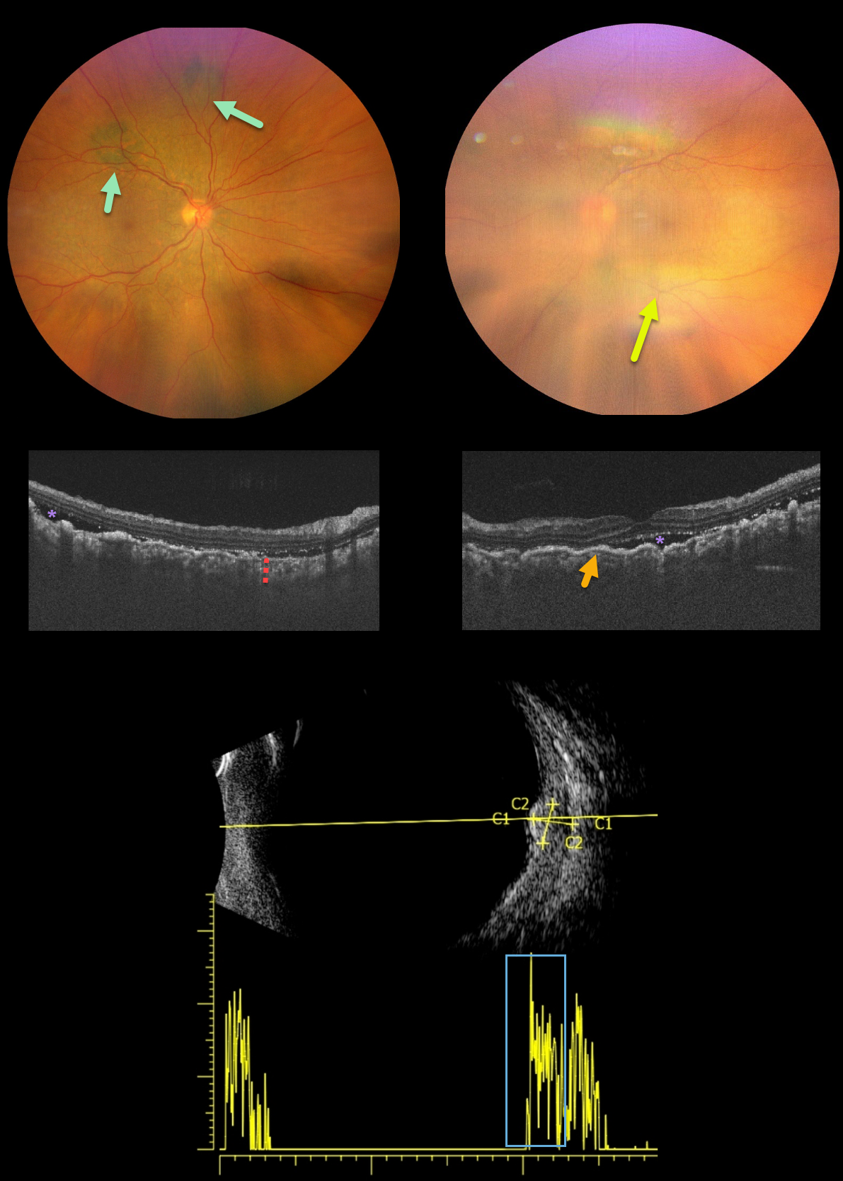
This is a 62YO female presented with bilateral vision loss over the past 3 months. Her vision was 20/200 OD and counting fingers OS compared to 20/20 OU at baseline. She was diagnosed with stage 4 ovarian cancer one month earlier.
New multifocal, elevated, pigmented choroidal lesions are noted superonasally in the right eye. View of the left fundus is limited due to a significant progressive cataract.
OCT reveals bilateral choroidal thickening with overlying folds and some subretinal fluid. B-scan ultrasonography through one of the choroidal lesions in the right eye shows moderate homogenous internal reflectivity.
Learning Points:
Initially described by Machemer in 1966, the five cardinal features of bilateral diffuse uveal melanocytic proliferation (BDUMP) include:
(1) multiple round or oval subtle red patches at the level of the RPE
(2) a striking pattern of multifocal areas of fluorescein angiographic hyperfluorescence from these lesions
(3) multiple slightly elevated pigmented and nonpigmented uveal melanocytic tumors with diffuse uveal thickening
(4) exudative retinal detachment
(5) rapidly progressive cataracts (Gass et al, Arch Ophthalmology 1990;108:527-533).
The thickened choroid and pigmentary changes are secondary to infiltration by benign uveal melanocytes, possibly in response to a primary visceral tumor (ovarian and uterine carcinomas in women, lung carcinoma in men) that produces melanocytic growth factors. Vision typically deteriorates to near blindness within a year of presentation. The serous detachments can resolve with plasmapheresis, although the overall prognosis is generally poor, with a mean survival of about 1 year from the time of ocular diagnosis.



