

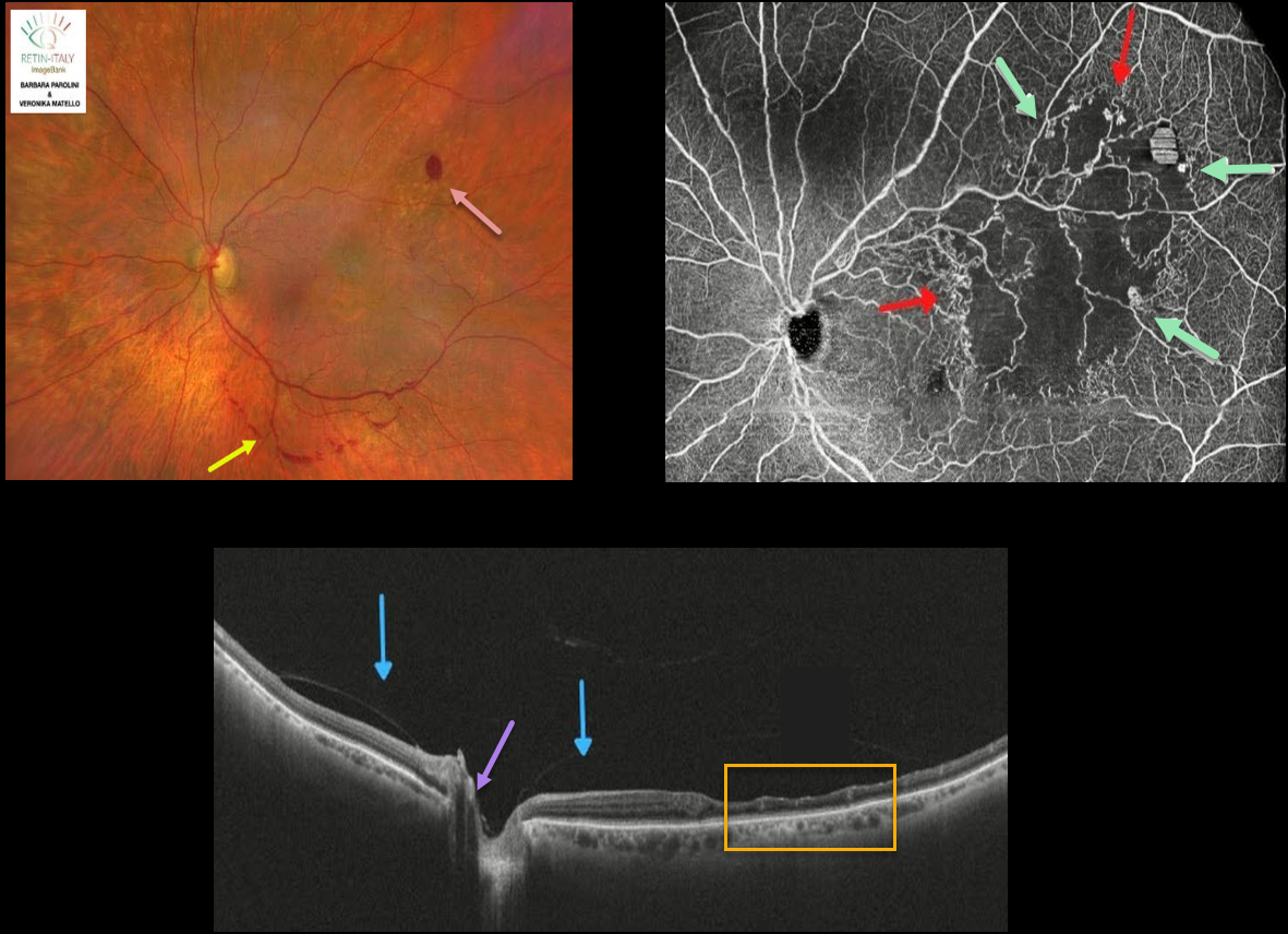
This 76YO female was referred for recent floaters. Wide-field color photography shows some retinal hemorrhages in the superotemporal retina, along with a band of preretinal blood below the inferotemporal arcade.
Wide-field SD-OCT shows inner retinal thinning within the area of occlusion temporally. The posterior hyaloid is detached over the posterior pole with a persistent adherence to the optic nerve.
Wide-field OCT angiography (OCTA) shows a broad area of capillary ischemia within the distribution of the branch retinal vein occlusion (BRVO), along with areas of retinal neovascularization.
Learning Points:
The endothelial cells in retinal neovascularization lack the tight junctions found in the normal retinal vasculature and therefore leak profusely on fluorescein angiography.
OCTA highlights retinal vessels not with a traditional fluorescing dye, but by detecting the movement of red blood cells within the vessel. This is why OCTA images offer beautiful detail but no leakage.
The role of OCTA versus traditional fluorescein angiography is still being determined, but growing evidence suggests it may provide similar clinical information without the need for dye injection (see Spaide et al, Progress Retinal Eye Research 2018;54;1-55).



