

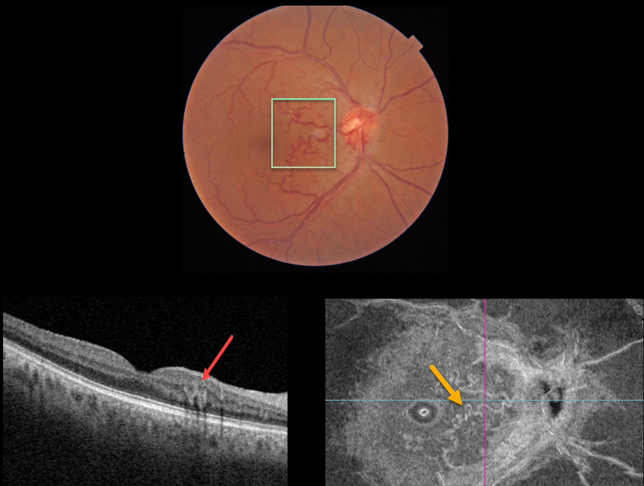
This 66YO male presented with an asymptomatic macular arteriovenous malformation (AVM). Vision was 20/30.
Triton imaging shows an AVM involving the nasal macula. Swept-source OCT shows that the abnormal vessels lie within the deep vascular complex. En face OCT beautifully displays the abnormal vasculature.
Learning Points:
Since about 25% of patients with AVMs can have intracranial venous anomalies, a brain MRI is usually recommended.
Our patient was asymptomatic, and his MRI and MRA were normal.



