

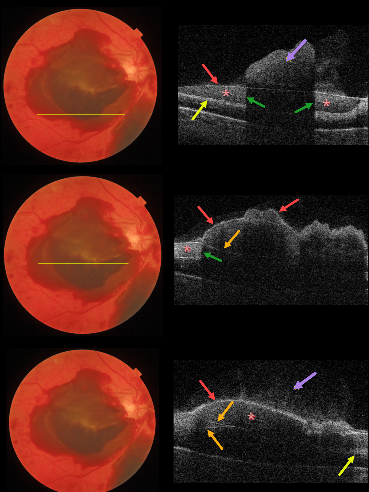
This 53YO female with type 2 diabetes presented with acute vision loss from previously undiagnosed proliferative diabetic retinopathy (PDR).
Color imaging shows multiple layers of preretinal blood. The blood is located above the neurosensory retina but is separated into several pockets due to posterior hyaloid splitting (vitreoschisis), rather than cleanly separating into a single plane.
Using the Triton swept-source OCT, we’ve taken four B-scans through different portions of the macula to highlight the complex anatomy of the preretinal blood.
Vitreous blood variably floats above the posterior hyaloid. The red arrows show the anterior edge of the posterior hyaloid. In some sections, the posterior hyaloid is split, separating the preretinal blood into several compartments.
Blood reflects and absorbs the OCT laser, making it difficult to see underlying retinal details (shadowing). The retinal nerve fiber layer is variably visible posterior to the blood.



