

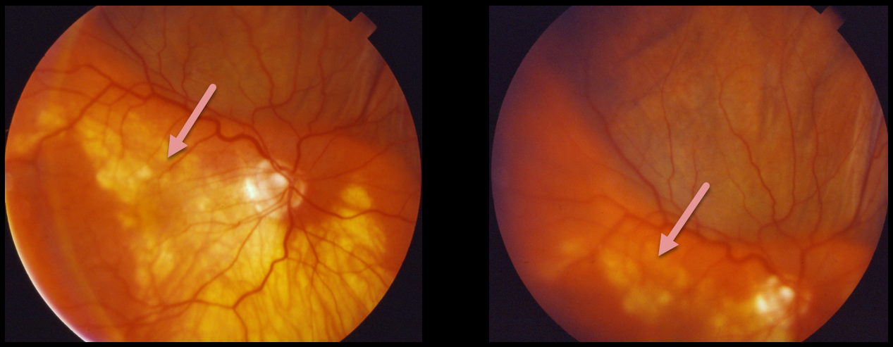
This patient presented with a pigmented, well-defined, and irregularly lumpy superior subretinal mass due to a suprachoroidal hemorrhage. This simulated a uveal melanoma.
Unfortunately, we don’t have any further information or images for this patient. However, we suspect that the blood spontaneously resolved with normalization of the fundus appearance.
Learning Points:
Patients presenting with suprachoroidal hemorrhage will usually have a history of recent intraocular surgery, trauma, or Valsalva. A clinical clue to this being a hemorrhage and not a melanoma includes the often somewhat lumpy and corrugated appearance, as in this patient.
See Chu and Green for a good review of suprachoroidal hemorrhage (Surv Ophthalmol 1999;43:471-486).



