

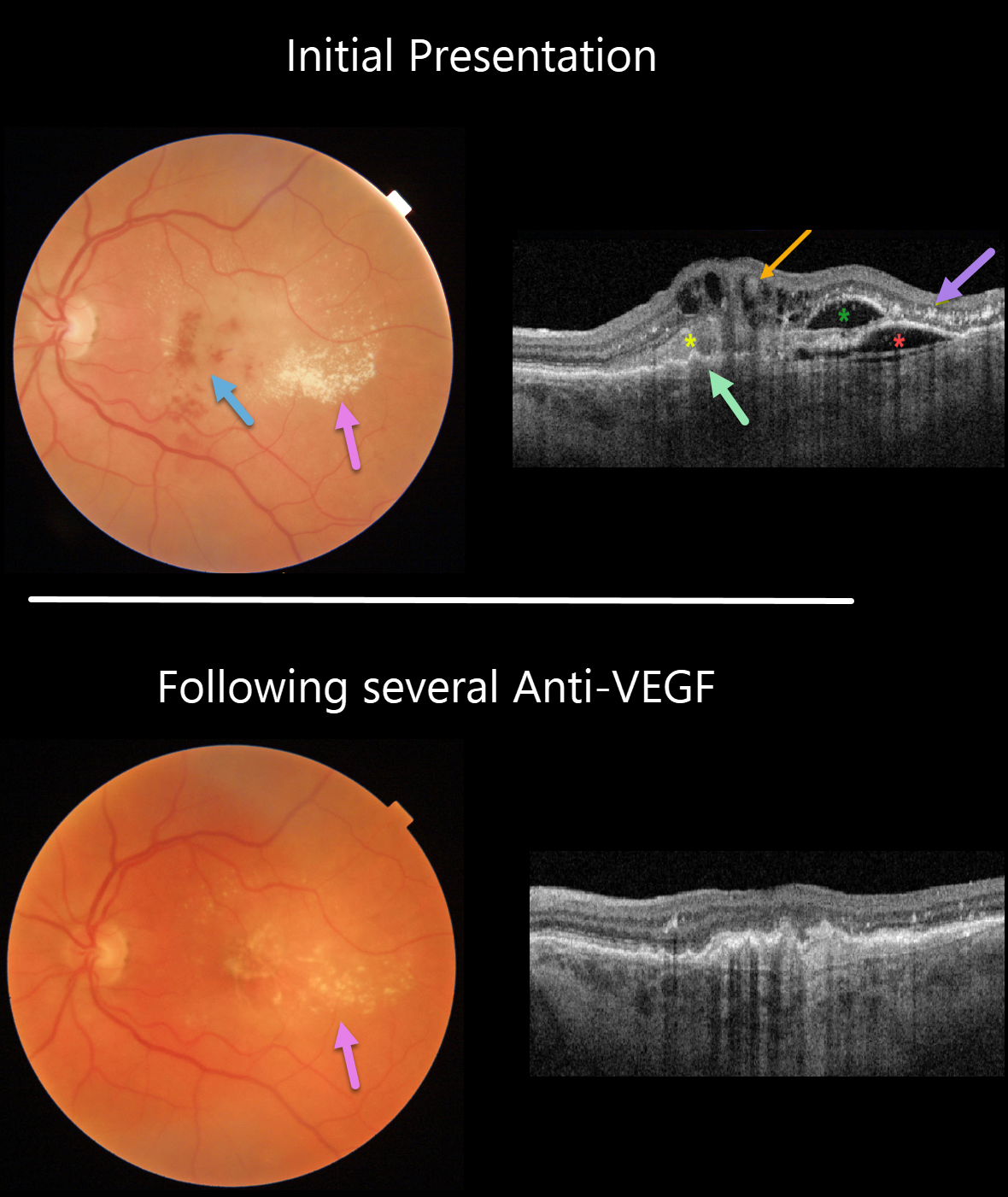
This 74YO man presented with counting fingers vision from previously untreated wet age-related macular degeneration (AMD).
Triton color imaging and swept-source OCT beautifully highlight this complex macular neovascularization (MNV). There are scattered inner and deep retinal hemorrhages, opaque fluid, and temporal lipid.
OCT shows an irregular shallow elevation of the RPE more nasally, possibly indicating a sub-RPE (type 1 MNV), with overlying subretinal hyperreflective material. More temporally, there is a more prominent area of RPE elevation that is more hyporeflective with an overlying pocket of intraretinal fluid vs a bacillary layer detachment. Finally, central inner retinal thickening and cysts are seen, indicating intraretinal neovascularization (type 3 MNV), also known as a retinal angiomatous proliferation (RAP) lesion. The lipid appears as hyperreflective clumps in the outer plexiform and outer nuclear layers.
The fluid and blood regressed, with significantly decreased lipid, after several monthly Avastin injections (image 2). Vision improved to 20/400.
Learning Points:
Unlike more typical age-related MNV that start either below (type 1 MNV) or occasionally above (type 2 MNV) the RPE, RAP lesions originate in the retina.
These incompetent vessels leak fluid and blood into the surrounding tissue. Their intraretinal location is a biomarker for RAP lesions.



