

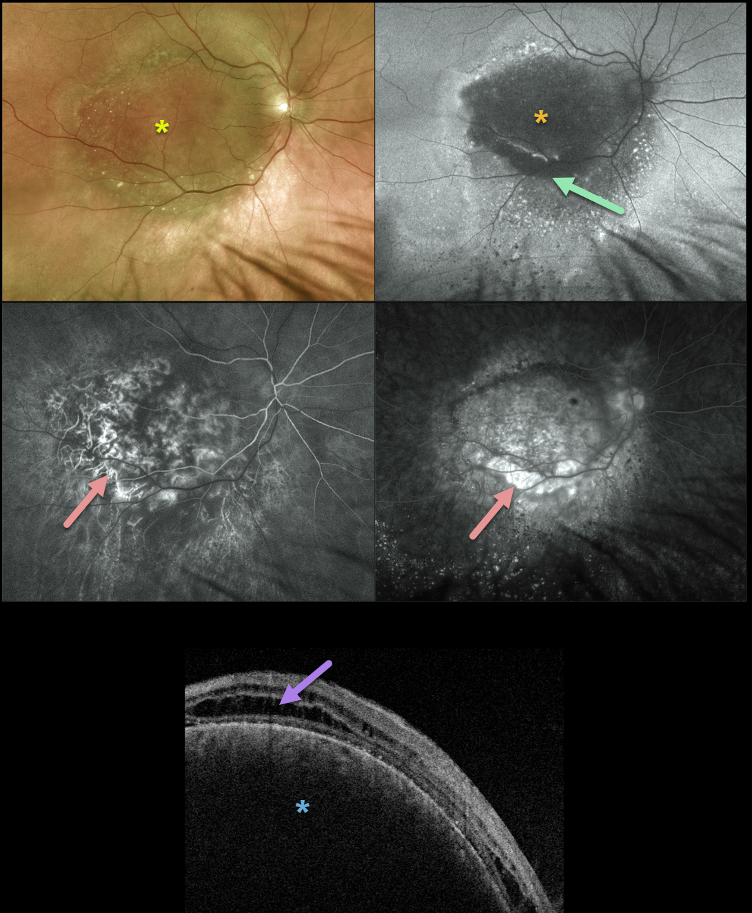
We have been following this 71YO male since 2014 with a choroidal hemangioma. Vision without correction was counting fingers, but improved with a +8.00 correction to 20/70. He tried a trial with a contact lens, but preferred to remain uncorrected due to visually disabling metamorphopsia.
The lesion has remained relatively stable over the years, and he continues to prefer observation over possible photodynamic therapy (PDT).
Multifocal Optos imaging on 8/3/21 shows a fairly stable tumor. Color imaging shows the hemangioma to be mostly orange-red with some more peripheral brownish coloration.
Fundus autofluorescence (FAF) reveals that the tumor is mostly hypo-FAF, indicating damage to the overlying RPE. There is a surrounding variable hyper- and hypo-FAF with a gutter of changes extending more inferiorly due to overlying exudative subretinal fluid.
Fluorescein angiography shows minimal leakage with mostly variable window-type defects. Triton swept-source OCT shows cystic edema overlying the markedly elevated choroidal tumor.
Learning Points:
Isolated choroidal hemangiomas are benign vascular lesions. Although they have no malignant potential, they can cause vision loss from exudation leading to subretinal and intraretinal fluid, which can be treated with PDT.



