

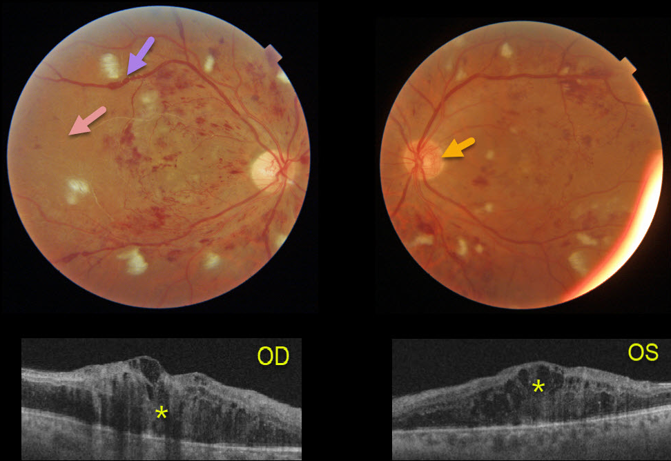
This 41YO male presented after a recent hospitalization for severe hypertension with vision of counting fingers OD and 20/60 OS.
The bilateral nerve fiber layer infarcts (cotton-wool spots) are more characteristic of hypertension than of diabetes.
Severe retinal hemorrhages and disc neovascularization are noted in each eye. The right eye, in particular, is severely ischemic, with Triton imaging showing a featureless, ischemic retina and white vessels, most pronounced in the temporal retina.
The venous sausaging is a clinical sign of severe ischemia, and when present, virtually always shortly precedes or is indicative of concurrent proliferative disease. Triton swept-source OCT shows severe bilateral center-involving diabetic macular edema.
He subsequently received multiple bilateral anti-VEGF injections followed by panretinal photocoagulation, with marked improvement of the central edema.



