

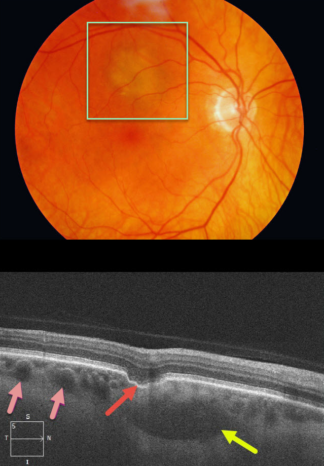
This patient was referred for an asymptomatic, slightly depigmented subretinal lesion in the superior right macula. OCT through this lesion reveals a focal choroidal excavation, choroidal pachyvessels, and probable loculated outer choroidal fluid.
Learning Points:
Focal choroidal excavation (FCE), originally described by Jampol et al in 2006, is likely part of the pachychoroid spectrum since it is usually associated with a thickened choroid and pachyvessels. The FCE can be observed, although symptomatic manifestations, including active central serous retinopathy or macular neovascularization, often require treatment.
Our patient also showed accumulation of fluid in the outer choroid. Spaide and Ryan noted this finding in about two-thirds of patients with central serous retinopathy (Am J Ophthalmology 2015;160;1211-1216). The presence of an FCE, pachyvessels, and loculated posterior choroidal fluid in our patient is intriguing and supports the association between pachychoroid disorders and FCE.



