

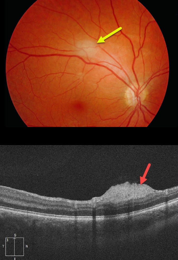
This patient with a known history of tuberous sclerosis was referred for an asymptomatic, slightly thickened whitish inner retinal lesion. OCT scanning showed an elevated, hyperreflective nerve fiber layer lesion.
Learning Points:
A retinal astrocytic hamartoma is a benign tumor composed of spindle-shaped fibrous astrocytes located in the nerve fiber layer, most commonly associated with tuberous sclerosis.
Tuberous sclerosis includes the classic triad of seizures, intellectual disability, and fibroangiomas, but patients can have lesions throughout the body, including the eyes, skin (ash-leaf spots), bones, nails, teeth, brain, heart, lungs, and kidneys.
Early in life, the inner retinal lesions appear fleshy white and are often multifocal and bilateral. Later in life, they show a more classic “mulberry appearance” with significant calcification. Rarely, the vascular component can cause intraretinal exudation requiring treatment.



