

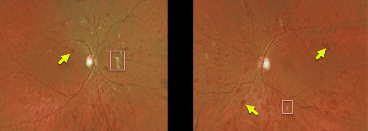
This patient with acute myeloid leukemia (AML) was referred by their oncologist for an eye examination following a failed allogeneic stem cell transplant due to graft versus host disease. Vision was 20/30 OD and 20/25 OS. His WBC was 14.7 (with 32% marrow myeloblasts), hemoglobin was 8.2, and platelet count was 23,000.
This patient has large, deep blot hemorrhages in the outer plexiform layer as well as scattered cotton-wool spots, indicative of leukemic retinopathy.
Learning Points:
These deeper hemorrhages would be unusual for the more common disorders seen in clinical practice (including diabetic retinopathy, retinal vein occlusions, and hypertensive retinopathy) and are usually indicative of blood dyscrasias, including anemia and hematologic malignancies. These deeper hemorrhages are also seen peripherally in ocular ischemia.



