

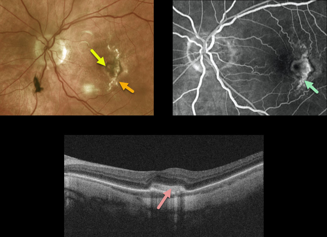
This 69YO female came in for a routine exam with a few months of vision loss in her left eye.
There is a new small area of subfoveal pigment adjacent to an old curvilinear scar more temporally. Although this temporal scar appeared to represent a choroidal rupture, she denied any prior trauma.
Axial length is 27.30 mm in this eye, nearly 1 mm greater than her fellow eye, indicating that this scar is likely a large and unusual lacquer crack.
Fluorescein angiography (FA) shows staining of the temporal scar and subfoveal pigment. An OCT B-scan through the subfoveal pigment shows a type 2 macular neovascularization (MNV) without fluid.
Learning Points:
Myopic MNV often presents as a pigmented subretinal lesion, often with minimal to no fluid due to the underlying thinned choroid.
In our experience, the presence or absence of new blood or new visual symptoms is more useful than the OCT, which usually shows no fluid, or FA, which often shows no leakage. Anti-VEGF injections were therefore recommended.



