

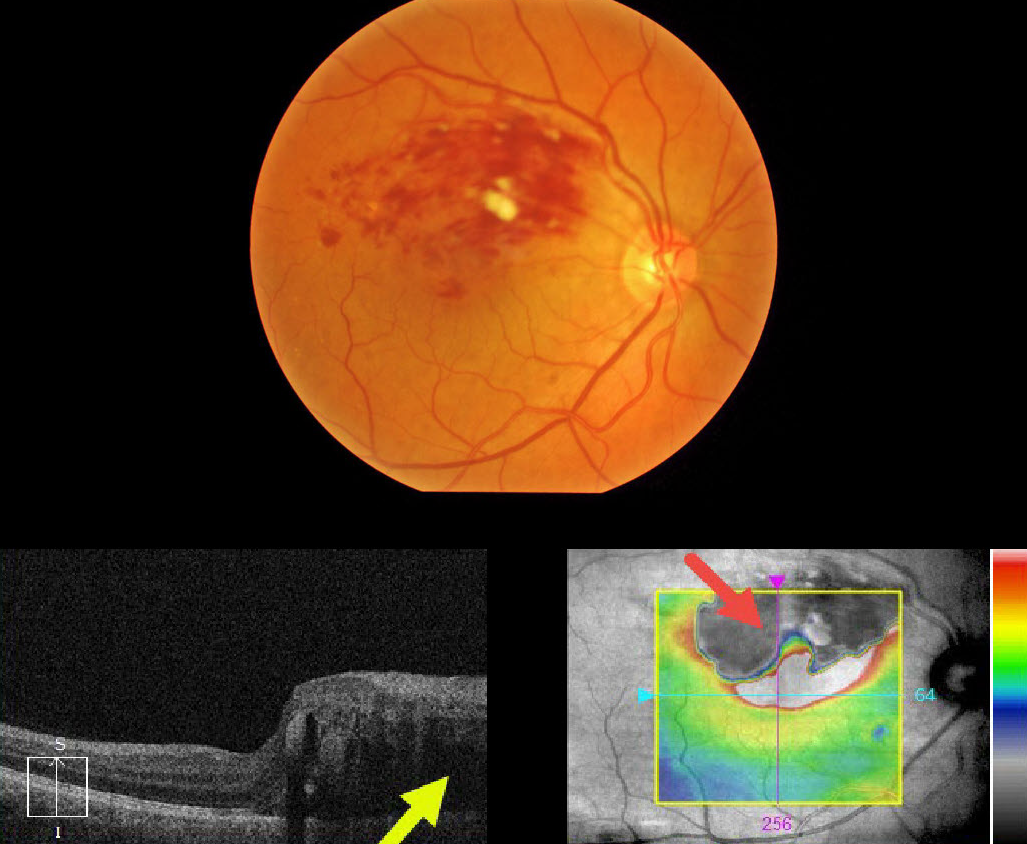
This 57YO male presented with 20/400 vision in his right eye from a superior macular branch retinal vein occlusion.
The OCT at first glance seems to just show superior macular edema extending through the macular center, but why is the superior retinal thickness map black, and what does that mean?
The retinal thickness map is supposed to represent the retinal thickness from the internal limiting membrane (ILM) to the tip of the outer segments just above the apical retinal pigment epithelium (RPE).
Our very expensive OCT machines usually accurately identify the ILM, but often don’t do such a great job of finding the RPE, especially when the OCT beam is attenuated as it passes through edematous retina or increased inner retinal reflectivity. In this case, the confluent inner retinal blood is highly reflective, causing posterior shadowing of the OCT signal.
Learning Points:
Intuitively, most of us would realize the black portion of the thickness map is an artifact, but now we know exactly what is going on. These artifacts are quite common if one looks for them.
The OCT cannot accurately locate the RPE signal and therefore incorrectly localizes it to the outer margin of the inner retinal blood.



