

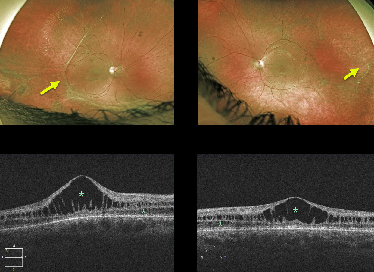
This 18YO male has classic findings for X-linked retinoschisis, including vitreous veils and bilateral foveal schisis. The macular schisis involves mostly the inner and outer nuclear layers, while peripherally it involves the nerve fiber layer (not pictured). Vision is 20/400 OD and 20/100 OS.
Learning Points:
X-linked retinoschisis is caused by a mutation in the RS1 gene, located on the X chromosome and encoding retinoschisin, a protein likely involved in retinal cell adhesion. Although it is usually transmitted as an X-linked recessive disorder in males, it can also affect females.
Over time, the macular schisis flattens with secondary atrophy. Vision usually levels off at about 20/100. Patients can develop spontaneous vitreous hemorrhage and rhegmatogenous retinal detachment from peripheral schisis.
Although there is no specific treatment, topical carbonic anhydrase inhibitors may reduce foveal thickness and possibly minimize foveal atrophy.



