

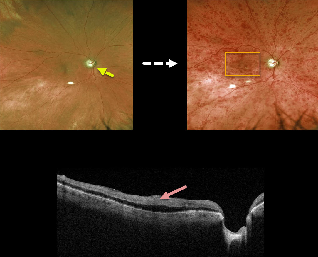
This 79YO male with a history of chronic open-angle glaucoma (COAG) presented with counting fingers vision from an acute nonischemic central retinal vein occlusion.
Even though his COAG was well-controlled with topical therapy, we felt that his glaucoma was a significant risk factor for the occlusion. A fundus photo from before the CRVO shows that significant glaucomatous cupping developed after the CRVO.
Triton swept-source OCT scanning shows marked inner retinal hyperreflectivity from the confluent macular blood.
Learning Points:
Patients with CRVO are at risk of vision loss due to macular edema or anterior segment neovascularization.
Macular edema is common in both non-ischemic and ischemic occlusions, although rubeosis is found almost exclusively with ischemic occlusions. Inner macular blood, as in this patient, can also cause vision loss.
Iris neovascularization with neovascular glaucoma develops in about two-thirds of patients with untreated ischemic occlusions. Rubeosis is treated initially with an anti-VEGF injection, followed by aggressive, complete panretinal photocoagulation.
Some patients will need a glaucoma tube or a cyclodestructive procedure if the IOP remains too high.



