

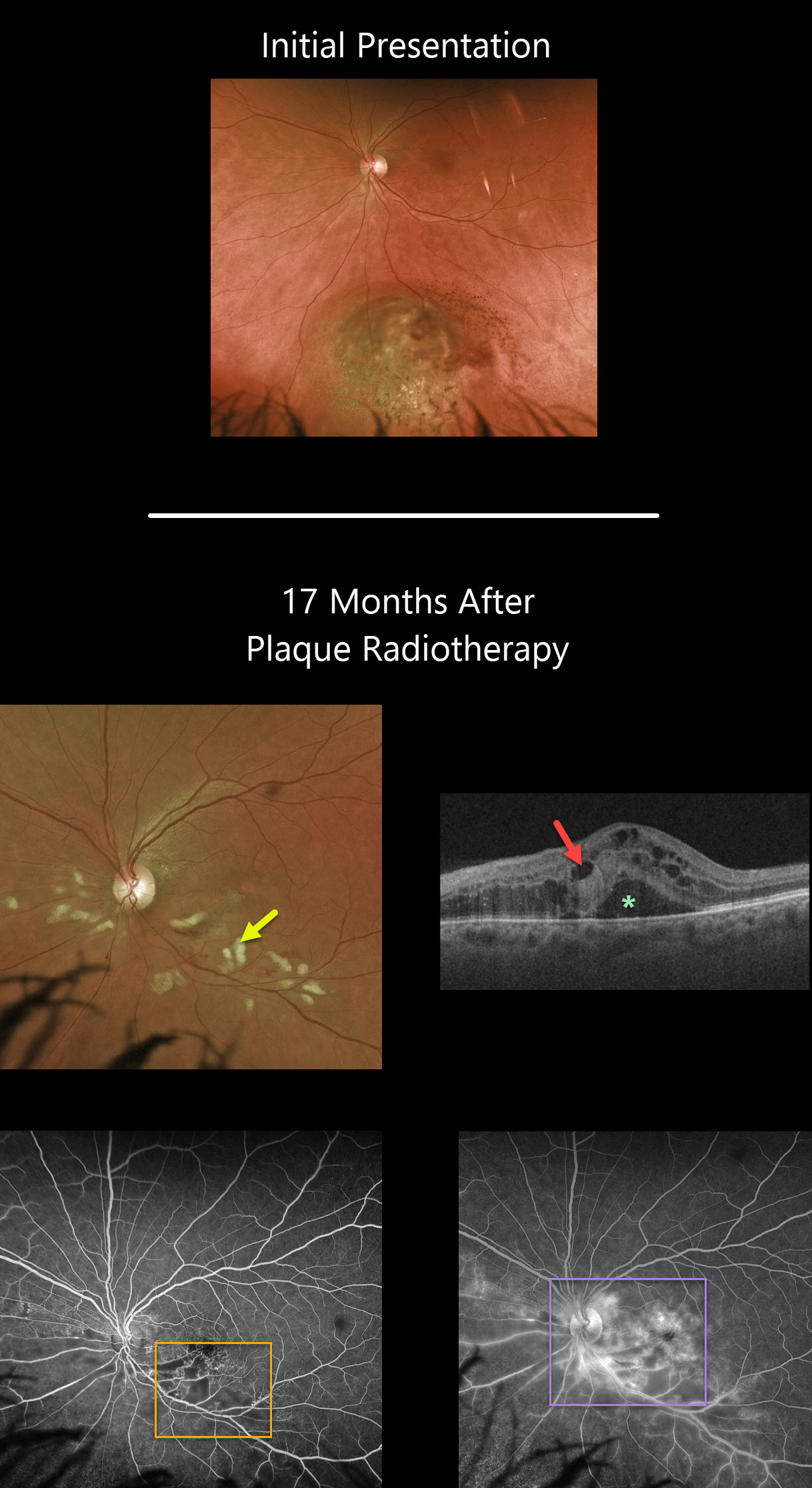
Seventeen months following plaque radiotherapy for a uveal malignant melanoma, this patient developed radiation retinopathy with scattered inner retinal hemorrhages and nerve fiber layer infarcts (cotton wool spots). Vision was counting fingers, although it had been 20/40 ten days following plaque therapy a year and a half earlier.
Cystoid macular edema with subretinal fluid is noted on Triton swept-source OCT. Fluorescein angiography shows areas of capillary dropout, telangiectasias, and hyperfluorescent leakage secondary to cystoid macular edema.
Learning Points:
Radiation retinopathy usually occurs 3 months to 3 years after external beam radiation (e.g., for facial or orbital cancers) or plaque radiotherapy. The ocular findings closely resemble those seen with diabetic retinopathy, as in our patient, who is receiving Avastin injections for the macular edema.



