

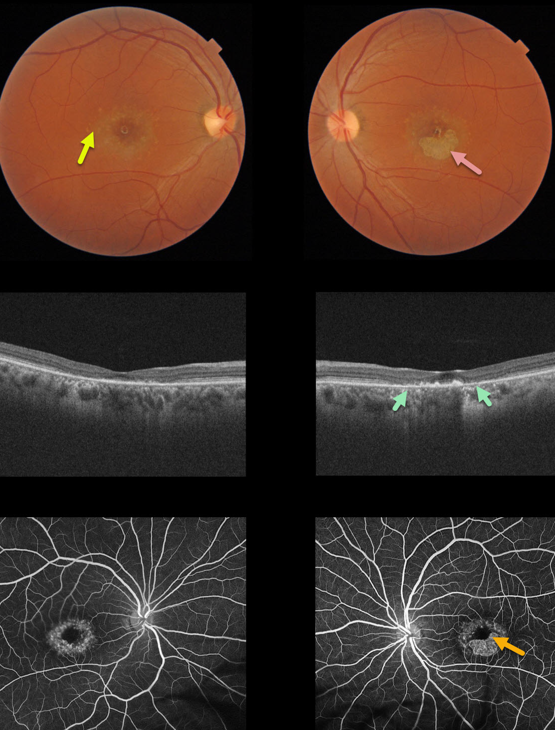
This 40YO female presented with vision of 20/400 in her right eye and counting fingers (CF) in her left eye due to classic findings from Stargardt’s disease, including perifoveal yellow subretinal flecks and variable outer retinal/RPE atrophy.
The OCT of her left eye best visualizes the bull’s-eye pattern of outer retinal and RPE atrophy. Loss of photoreceptors is evident as the outer plexiform layer (OPL) descends towards the RPE, eventually resulting in fairly complete atrophy of the outer retina and RPE.
Fluorescein angiography (FA) characteristically shows a ‘dark choroid’ from the lipofuscin-engorged RPE blocking the choroid (not present in this case).
Learning Points:
Stargardt’s disease is an autosomal recessive disorder caused by a mutation in the ABCA4 gene. The ABCA4 protein is located in the photoreceptor outer segments and is involved in the recycling of 11-cis-retinal.
Mutation of this gene causes accumulation of lipofuscin in the RPE, eventually leading to photoreceptor and RPE degeneration. Other ABCA4 disorders include fundus flavimaculatus and cone-rod dystrophy.



