

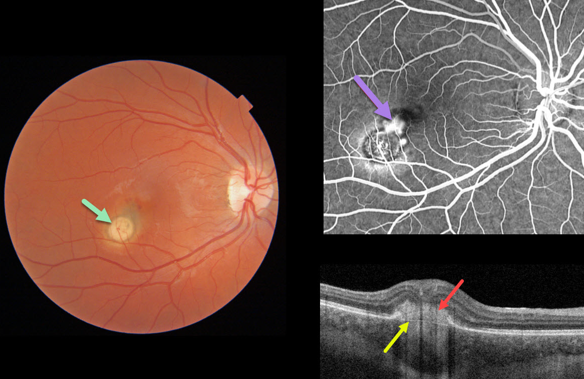
This 22YO female presented with 20/400 vision from a macular neovascularization (MNV) associated with a choroidal granuloma.
The Triton image shows a focal yellow-white nodular subretinal lesion with adjacent subretinal pigment and blood along its foveal edge.
Swept-source OCT through the MNV shows the hyperreflective MNV above Bruch’s membrane. Fluorescein angiography shows leakage from the MNV.
Learning Points:
Choroidal granulomas usually appear as solitary, creamy white, round subretinal lesions. They are hyporeflective on OCT imaging (Invernizzi et al, Retina 2015;35:525-531). Causes include sarcoidosis, tuberculosis, and VKH. They can occasionally cause exudation that responds to anti-VEGF injections or photodynamic therapy.



