

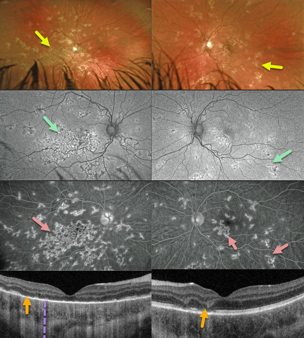
This 12YO boy was referred for asymptomatic retinal changes. Vision was counting fingers OD and 20/25 OS.
Optos imaging shows patchy inactive chorioretinal scarring extending from each macula to the mid-periphery, especially confluent in the right macula.
There is variable hyper- and hypo-autofluorescence, and fluorescein angiography shows areas of staining. Triton swept-source OCT shows variable outer retinal loss with fairly intact RPE associated with increased choroidal transmission.
Our presumed diagnosis was relentless placoid chorioretinitis. Since all the funduscopic findings appeared inactive, we are currently observing him without treatment.
Learning Points:
Relentless placoid chorioretinitis has clinical and angiographic features resembling both serpiginous choroiditis and acute posterior multifocal placoid pigment epitheliopathy (APMPPE).
Relentless placoid demonstrates the multifocal lesions of APMPPE, with more meandering, less patchy, serpiginous lesions.
Additionally, these patients will have recurrent inflammation typical of serpiginous but not APMPPE. They may also develop macular neovascularization, which is more typical of serpiginous.



