

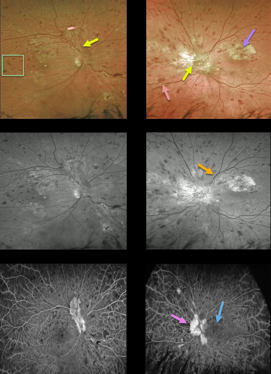
This 40YO man presented with bilateral proliferative diabetic retinopathy (PDR). Vision was 20/60 OD and 20/200 OS.
Optos color and green channel imaging show prominent bilateral fibrovascular disc neovascularization, obscuring the left disc.
A few smaller areas of more peripheral neovascularization are present nasally in the OS. Severe retinal hemorrhages are noted in all quadrants; the retinal veins are beaded OU superiorly, along with some perivascular lipid superotemporally OD and nasally OS. The retinal vascular abnormalities are best highlighted with the green channel image.
There also appears to be an area of fibrosed neovascularization in the superior left macula. However, these findings are visible beneath the retinal vessels, indicating they are subretinal and composed of lipid. Lipid is usually angiographically invisible but, when dense, can obscure the underlying choroidal fluorescence, as in this case.
Fluorescein angiography shows leakage from the neovascularization, diffuse vascular staining, and mostly peripheral retinal nonperfusion.



