

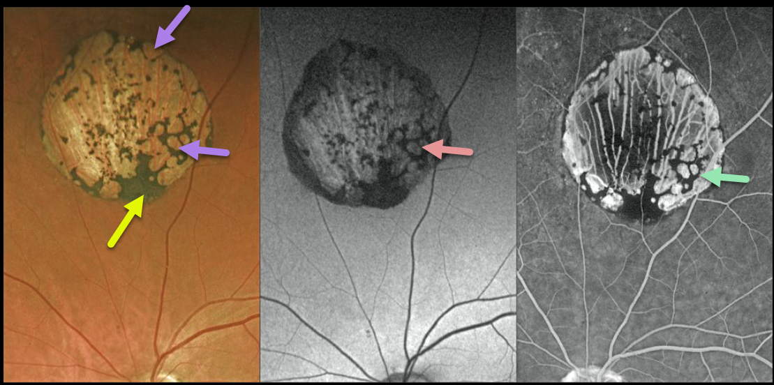
This is a gorgeous multimodal Optos image of a classic solitary congenital hypertrophy of the RPE (CHRPE). The borders are well-demarcated with a variable coal-black coloration.
With age, they usually develop multiple hypopigmented lacunae that tend to enlarge over time. These lacunae demonstrate hyperautofluorescence on fundus autofluorescence (FAF), whereas on fluorescein angiography (FA), they appear as hyperfluorescent window defects.
The CHRPE lesions can also enlarge with time, but carry virtually no malignant potential.
Learning Points:
Histologic RPE hypertrophy presents in several ways. Typical isolated CHRPE lesions and bear tracks are benign ocular findings and require no systemic evaluation. Only the multiple, small comet-shaped lesions are part of the familial polyposis spectrum.



