

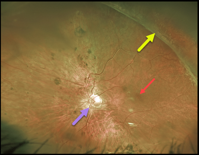
For over 20 years, we’ve been following this 39YO male with stable regressed retinopathy of prematurity in his left eye. Vision is NLP OD and 20/80 OS.
Optos imaging shows that the macular center is dragged temporally due to vitreoretinal traction from regressed neovascularization.
The macular center is usually about 2 disc diameters temporal to the edge of the optic nerve, and our patient’s is about 3 disc diameters. This dragging is usually accompanied by a straightening of the temporal arcades.
However, in our patient, this is masked by situs inversus, a congenital abnormality in which the retinal vessels exit the optic nerve with a nasal bend before coursing temporally.
Learning Points:
These outcomes can be minimized through aggressive neonatal screening and treatment with laser photocoagulation and anti-VEGF agents.



