

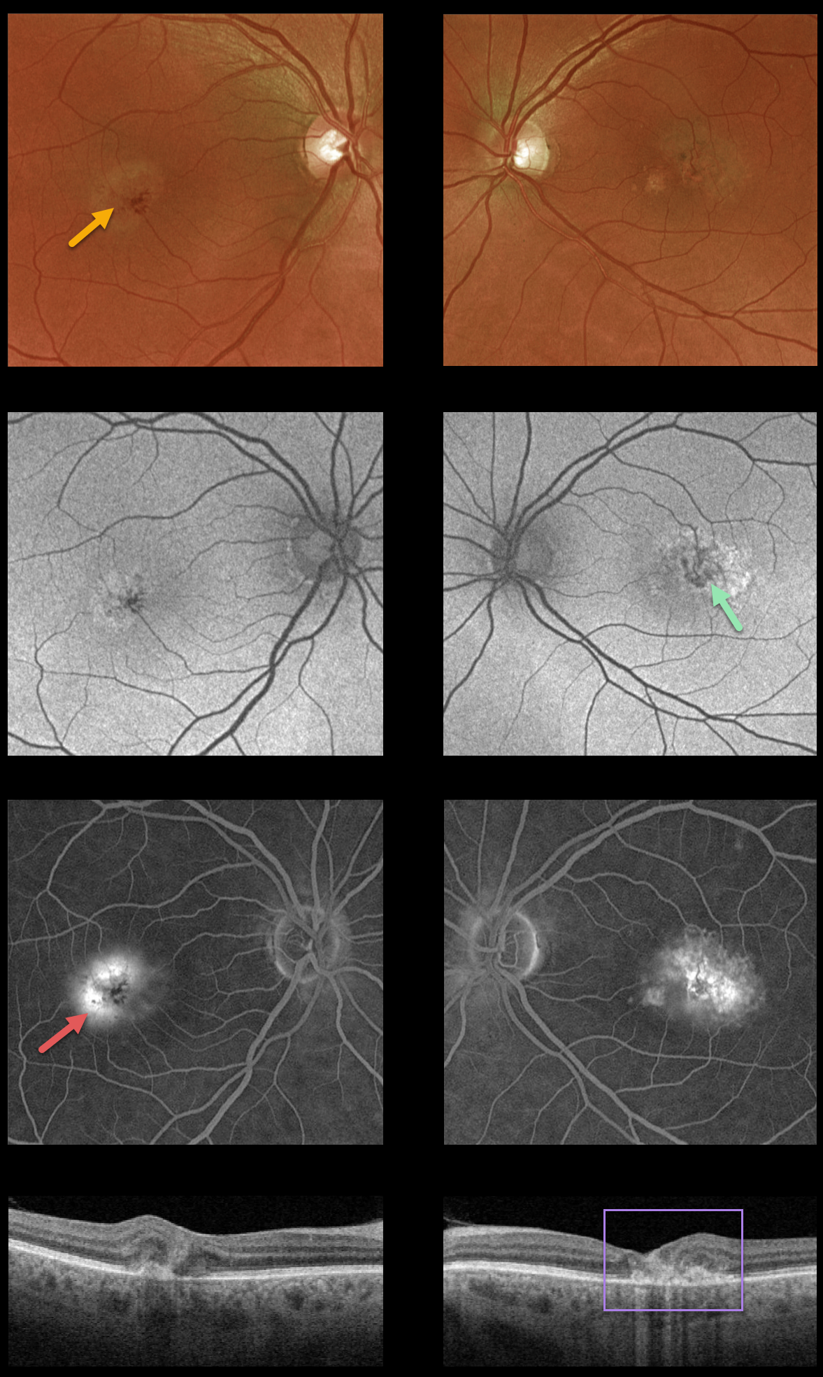
This 55YO male presented with macular telangiectasia type 2 (MacTel2). He was visually asymptomatic, and vision was 20/40 OD and 20/50 OS.
This case highlights various multimodal imaging findings. The foveal retina shows a whitish discoloration, most likely due to retinal opacification resulting from Müller cell dysfunction.
There are variable macular hyper- and hypofAF abnormalities, with angiographic staining and leakage. OCT scanning shows mostly temporal foveal outer retinal atrophy and disorganization.
Characteristic MacTel2 features absent in our patient include refractile inner retinal ‘crystals’ (thought to be Muller cell footplates, similar to the refractile dots found in retinoschisis) and coarse black clumps of intraretinal pigment migration.
Learning Points:
Originally described by Gass (Arch Ophthalmology 1982;100:769-780), MacTel2 is a neurodegenerative disorder, most likely originating from Muller cell dysfunction. The term telangiectasia is misleading because the funduscopic findings are mostly nonvascular.
Although much remains to be learned about its pathophysiology and there is no treatment for the underlying disease, secondary macular neovascularization can be successfully treated according to age-related macular degeneration protocols.



