

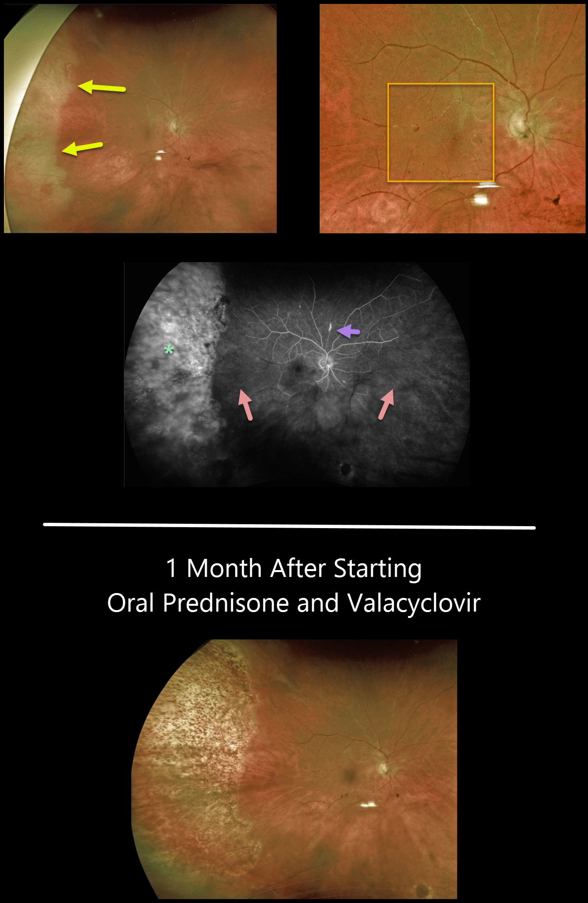
This 80YO female presented with a sudden onset of floaters while battling 2 months of persistent iridocyclitis from Herpes zoster ophthalmicus.
There was increased anterior uveitis, along with new peripheral temporal retinitis. Despite significant macular ischemia on clinical examination, vision was 20/60. Our patient’s posterior involvement was likely seeded from her anterior segment.
Optos fluorescein angiography shows diffuse staining of the peripheral retina, extensive nonperfusion throughout the posterior pole, and a few scattered areas of focal staining vasculitis.
She was started on high-dose oral prednisone and valacyclovir. One month later, vision improved to 20/40, and the retinitis resolved with secondary chorioretinal scarring.
We are slowly tapering her steroids and plan to leave her on low-dose valacyclovir as prophylaxis against recurrent infection in her right eye, as well as infection in her normal left eye.
Learning Points:
Acute retinal necrosis syndrome (ARNS) is most commonly caused by the Herpes zoster virus. ARNS, which usually occurs in immunocompetent individuals, is a rapidly progressive panuveitis with hemorrhagic and ischemic unilateral (ARNS) or bilateral (BARNS) retinal necrosis that typically begins in the retinal periphery.
ARNS is often a devastating and blinding infection due to the high risk for retinal detachment as well as macular and optic nerve involvement.
The latest meta-analysis argues for systemic antiviral therapy and prophylactic vitrectomy, with uncertain benefits for prophylactic laser photocoagulation or adjunctive intravitreal antivirals (see Zhao et al, Retina 2021;41:965-978).



