

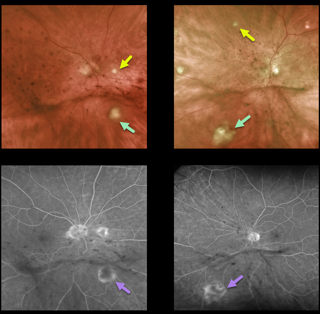
This 75 yo retired physician presented with decreased vision and floaters for about 8 weeks. Vision was 20/25 OD and 20/70 OS. Anterior segments were normal except for mild nuclear sclerosis.
There are multifocal areas of yellow-white chorioretinal inflammation spreading into the overlying vitreous, along with clumps of inflammatory deposits on the back of each hyaloid. Fluorescein angiography reveals early blockage with late surrounding staining of these inflammatory lesions.
He gave a history of non-Hodgkin’s lymphoma, which was in remission. He still had a port in place despite having completed chemotherapy treatments. He was also being treated with oral prednisone and azithromycin for organizing pneumonia and had active oral candidiasis.
Based on his clinical findings, immunosuppression, chemotherapy port, and oral thrush, we suspected bilateral candidal endophthalmitis, although we were also concerned about an atypical presentation for intraocular lymphoma.
Diagnostic vitrectomy was performed. Cytology was negative for lymphoma, and Gram stain and fungal cultures were also surprisingly negative. Blood cultures were also negative.
Despite the negative workup, we still felt that fungal endophthalmitis was the most likely diagnosis and started oral fluconazole. Since the port was no longer in use, this was removed and did, in fact, grow out Candida. The intraocular inflammation subsequently improved.
Learning Points:
In many cases of endophthalmitis, no infectious agent is identified, and often the diagnosis is made clinically. Empiric treatment often includes intravitreal antibiotics. In this case, despite negative blood cultures and vitreous biopsy, our clinical suspicion ultimately led to the correct diagnosis and treatment.



