

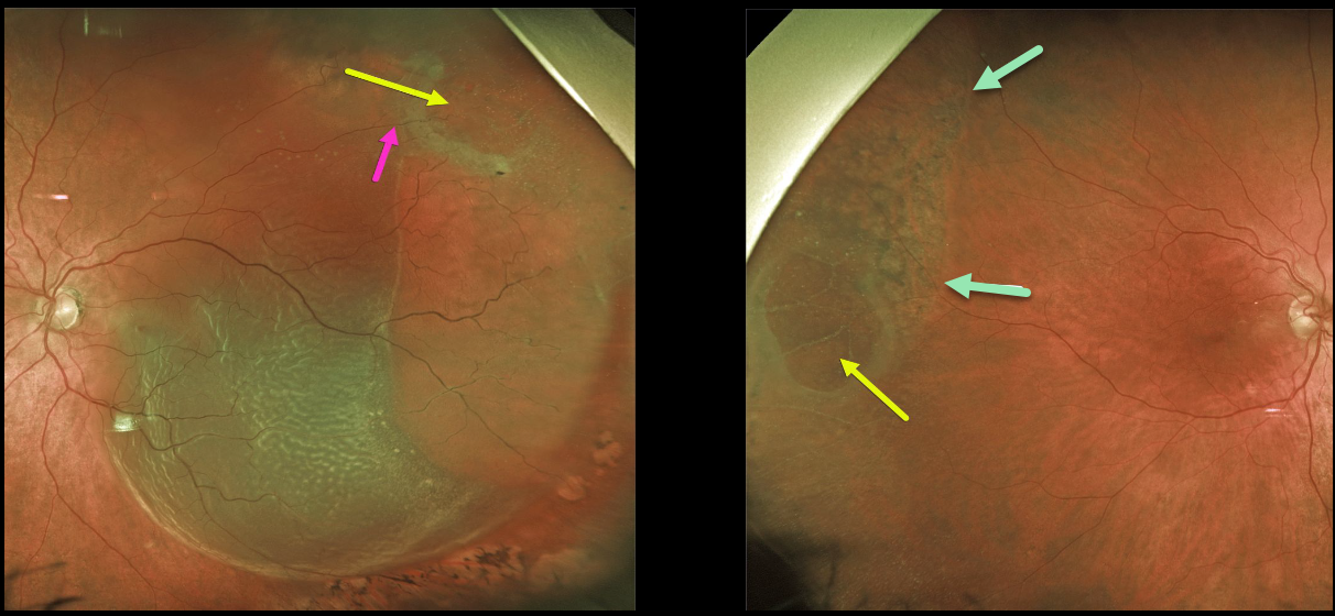
This 72YO male presented with an acute macula-off retinal detachment (RD) in his left eye.
Optos imaging shows that this is not a typical rhegmatogenous RD since an outer layer hole (OLH) is noted superotemporally. A retinal vessel can be traced over the OLH, indicating that this is not a full-thickness defect.
The patient also has degenerative retinoschisis with an OLH in his right eye. A shallow subclinical schisis detachment is likely present due to the pigmentary changes throughout the schisis cavity.
Prophylactic treatment is not recommended for this eye, but we are carefully following him, as he does have a small risk of developing an RD here as well.
Learning Points:
This detachment in the left eye does not have a typical full-thickness break, but is caused by degenerative retinoschisis.
Numerous tiny inner-layer holes, along with the visible large OLH, essentially created a full-thickness retinal defect, giving the liquid vitreous access to the subretinal space. The detachment was successfully repaired with vitrectomy surgery.



