

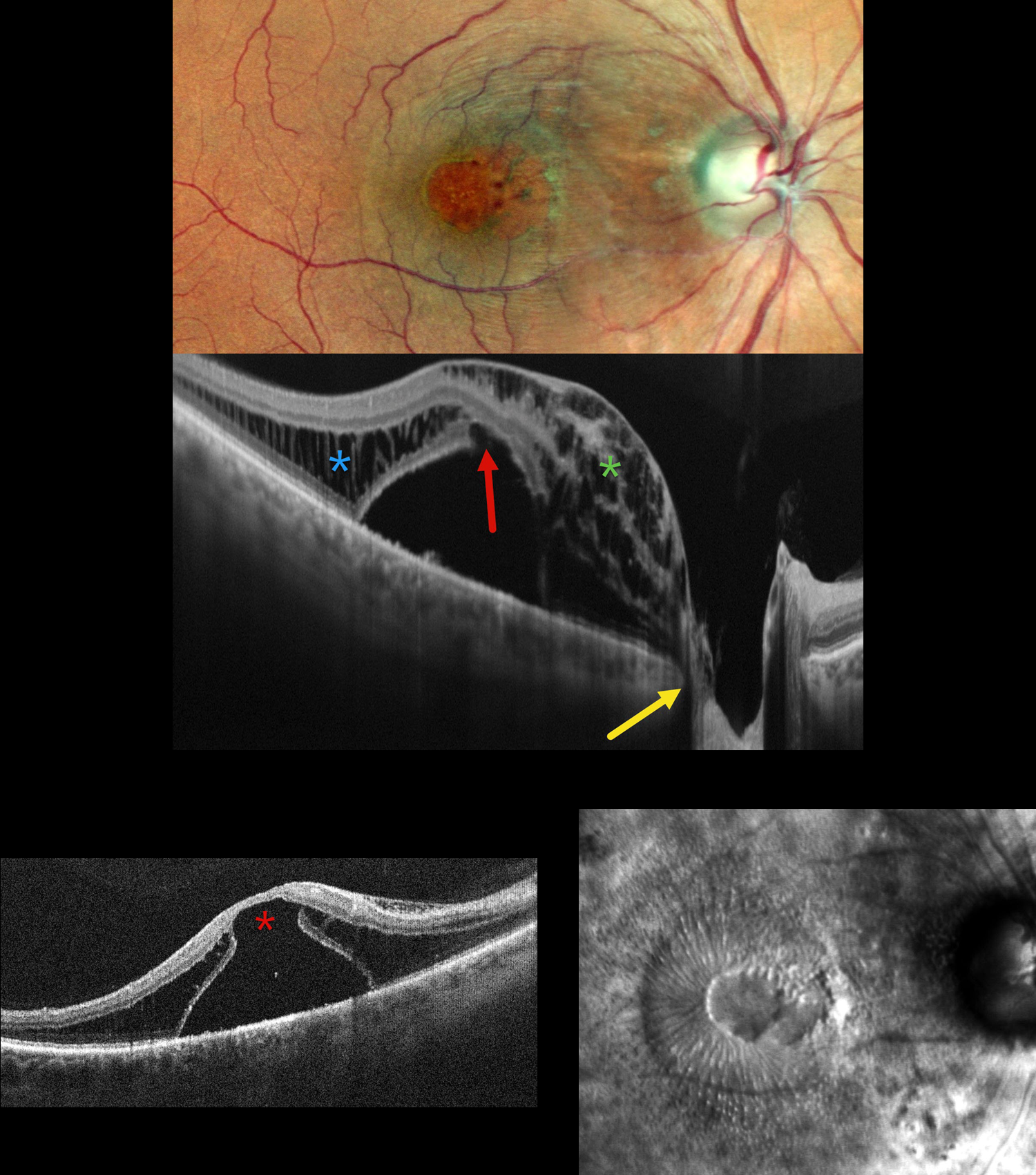
This 50YO male presented with 1 year of decreased vision in his right eye. He had been treated elsewhere with intravitreal injections for cystoid macular edema. Vision was 20/200 OD and 20/20 in his normal OS.
Pseudocolor SLO imaging shows a possible serous macular detachment with a large macular pseudohole. A possible pit is noted along the temporal aspect of the optic nerve. OCT scanning shows an elevated serous detachment. An outer-layer macular hole is noted along the posterior aspect of the fovea, along with full-thickness cystic retinal edema nasally and outer nuclear layer cystic edema temporally. The subretinal fluid directly communicates with the optic nerve pit. Retro mode reveals a unique view of the macular pathology, with the central outer macular hole and radiating fluid.
Learning Points:
An optic nerve pit is a rare (about 0.1% prevalence) congenital anomaly thought to be caused by incomplete closure of the optic fissure during gestation. Pits are most often located within or along the inferior-temporal disc margin and usually appear as a gray depression. While most optic pits are asymptomatic, they can cause vision loss by forcing fluid into the inner retina, outer retina, and, ultimately, the subretinal space (optic pit maculopathy). The source of the fluid remains a subject of ongoing debate, with opinions ranging from the vitreous to the subarachnoid space.
Vitrectomy surgery was discussed, and the patient elected for observation unless his symptoms worsened.



