

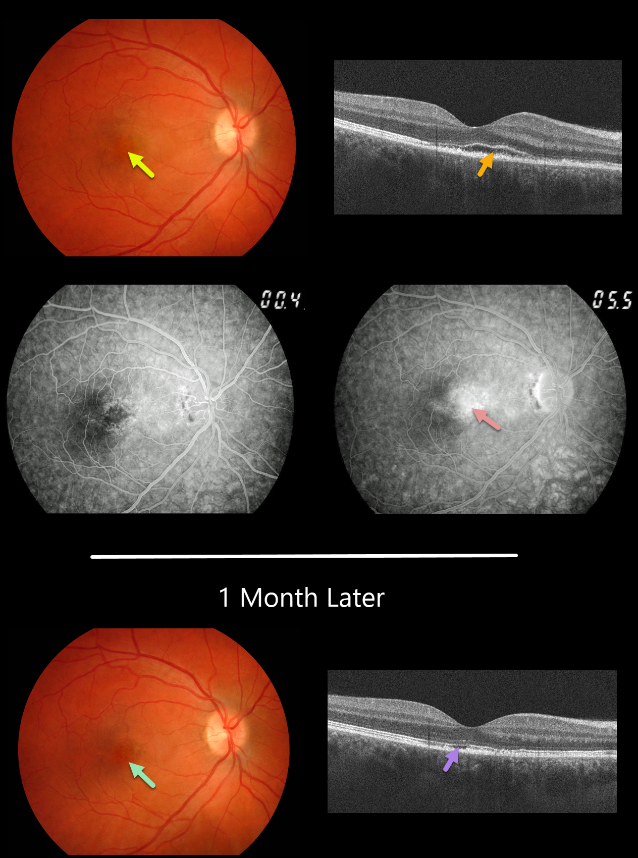
This 29YO male presented with a 1-week history of acute blurred vision in his right eye. Vision was 20/40 OD.
Fundus photography and fluorescein angiography reveal pigment mottling and late-stage leakage. OCT scanning shows an irregularly elevated ellipsoid zone with underlying outer segment disruption vs subretinal fluid and a thickened RPE.
We felt the patient had acute idiopathic maculopathy (AIM) and observed him without treatment.
One month later, the outer fluid resolved with residual irregularities in the outer retinal and RPE bands on OCT and an area of macular hyperpigmentation surrounded by a rim of pigment loss.
Learning Points:
Originally described by Yannuzzi et al. in 1991 (Arch Ophthalmology 1991;109:1411-1416), AIM is an idiopathic inflammation of the outer retina, RPE, and inner choroid, usually found in young people and often following a viral illness. It is usually self-limited, resolving within several weeks with significant visual improvement. The appearance of a bull’s-eye lesion, as occurred in our patient, is classic for this disease.



