

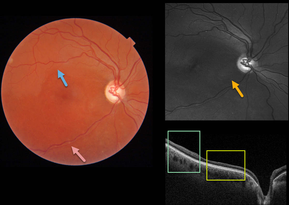
This healthy 30YO female was referred for poor vision in her right eye. She mentioned losing vision in this eye during high school, but couldn’t provide any further details. She also gave a history of hearing loss, which had never been evaluated. Vision was HM OD and 20/30 in her normal left eye.
Triton imaging shows faint retinal whitening in the superior macula. The retinal arterioles are narrowed and somewhat sheathed inferiorly.
Swept-source OCT shows normal inner retinal architecture temporally, with inner retinal atrophy and disorganization of the normal architecture more nasally. Right inferotemporal nerve fiber loss is quite dramatic on Optos green imaging.
We diagnosed her with a resolved inferior branch retinal artery occlusion (BRAO), although this may represent Susac’s syndrome. The involved retina appeared redder than the uninvolved superior retina, accounting for the whitish superior retina noted above.
Subsequent work-up, including hypercoagulable labs and MRI, was negative. She denied prior intravenous drug use. To complete the workup, we will be obtaining carotid Doppler and cardiac ultrasonography.
Learning Points:
Susac’s syndrome, also known as retinocochleocerebral vasculopathy, is a rare microangiopathic disease characterized by a triad of encephalopathy, sensorineural hearing loss, and branch retinal artery occlusions with a predilection for younger females.
In the presence of Susac’s syndrome, MRI findings should show corpus callosum involvement. However, the full triad commonly takes months to develop, complicating diagnosis.



