

This 23YO male initially presented with 5 days of bilateral blurred vision and stabbing pain in his right eye. Vision was 20/400 OU.
Panuveitis was noted bilaterally, with moderate granulomatous anterior uveitis, moderate vitreous cells, macular vascular occlusion, and possible vasculitis (not shown).
Review of symptoms revealed occasional headache, lack of genital ulcers, but was positive for an occasional oral ulcer. Subsequent laboratory workup was negative, including ANCA, ANA, rheumatoid factor, ESR, FTA-ABS, RPR, QuantiFERON-TB Gold, Lyme, HLA-B27, and lumbosacral spine x-ray.
He was started on oral prednisone and topical steroid drops. He was then lost to follow-up.
When seen nearly a year later, vision was CF OD and 20/400 OS with significant panuveitis as before.
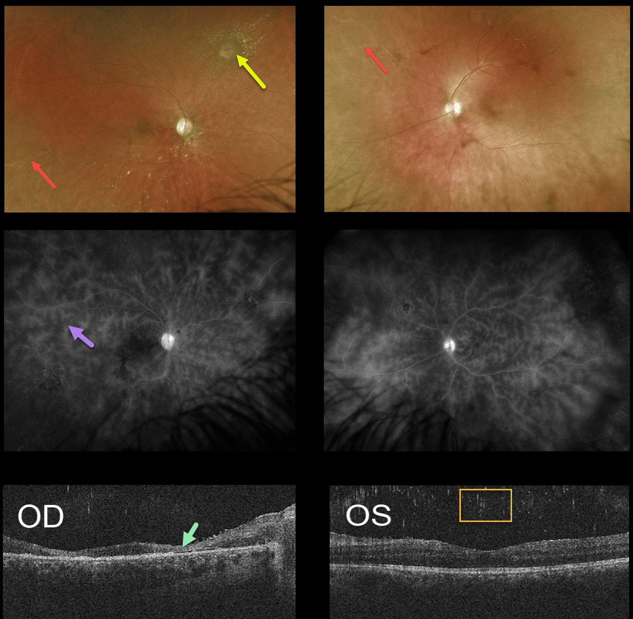
Optos imaging shows bilateral peripheral ischemia along with a whitish area of possible retinitis with surrounding lipid in the superonasal midperiphery of the right eye.
Fluorescein angiography shows diffuse vasculitis. OCT scanning shows severe retinal atrophy OD with bilateral vitreous cells.
Brain MRI revealed an enhancing lesion within the brainstem, and neurosurgery ruled out a malignancy. He was then referred to a neuro-immunologist. Further testing for infectious etiologies such as toxoplasmosis, toxocariasis, and Whipple’s disease was all negative. He then developed a 6th nerve palsy along with ataxia and was admitted to the hospital.
A combination of specialists, including neuro-immunology, rheumatology, and gastroenterology, felt there was a high probability of Behcet disease. The patient improved with a long course of oral prednisone and was slowly tapered as he was started on azathioprine.
Due to his severe peripheral ischemia and poor compliance with follow-up, he was treated with prophylactic scatter laser photocoagulation to prevent proliferative complications.
Learning Points:
Behcet disease is a systemic vasculitis that includes the classic triad of oral ulcers, genital ulcers, and ocular disease. There are no specific tests to confirm the diagnosis of Behcet’s.
The diagnosis is clinical and includes 1) recurrent oral ulcerations (aphthous or herpetiform) at least three times in one year AND 2) any two of the following: recurrent genital ulcerations, eye lesions (uveitis or retinal vasculitis), skin lesions (erythema nodosum, pseudofolliculitis, papulopustular lesions, acneiform nodules) found in adult patients not being treated with corticosteroids, and a positive “pathergy test” read by a physician within 24-48 hours of testing.
Anterior segment findings include uveitis and a sterile hypopyon. Posterior segment findings can be blinding and include an ischemic hemorrhagic retinal vasculitis. Vascular occlusions can cause optic atrophy, retinal neovascularization, and vitreous hemorrhage.
Treatment for the underlying uveitis and vasculitis is complex and includes topical/systemic corticosteroids, as well as immunomodulatory medications. Ocular neovascularization can be treated with anti-VEGF injections and scatter laser photocoagulation.



