

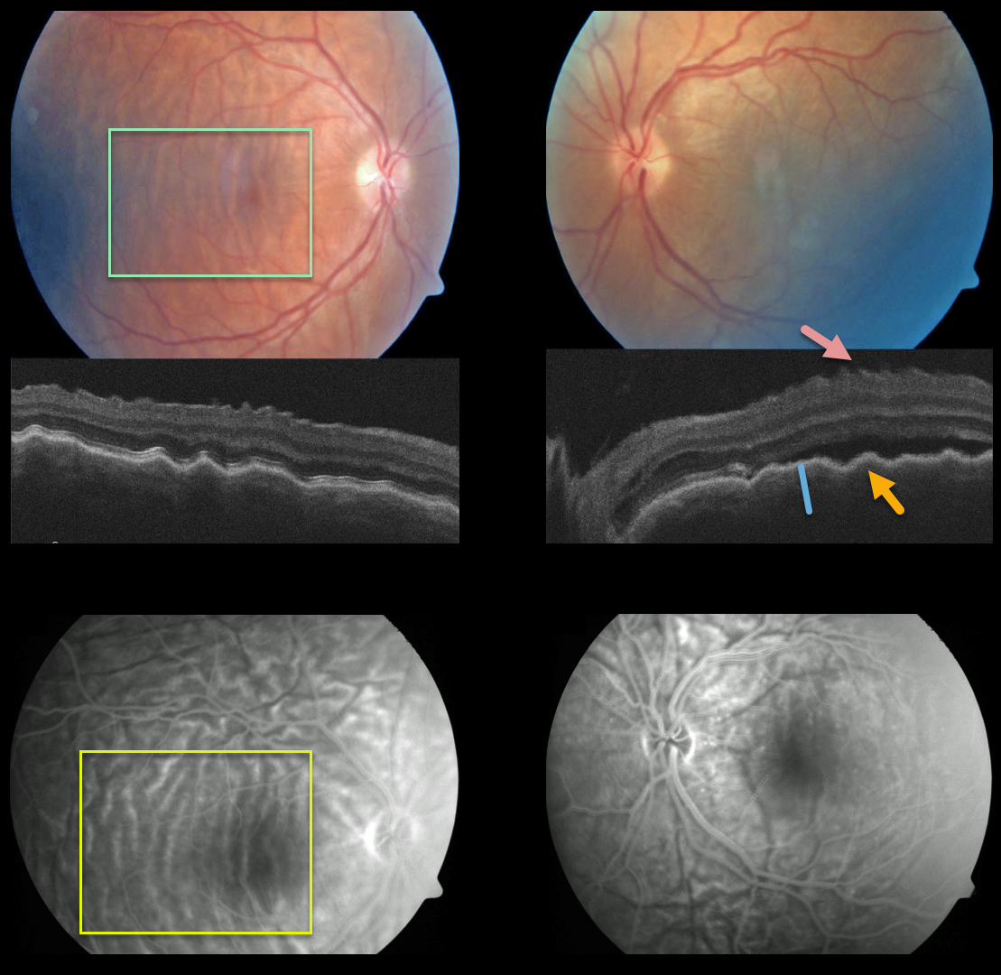
This patient presented with bilateral posterior pole chorioretinal folds, confirmed on OCT as inner retinal folds, outer retinal/RPE folds, and thickened choroid, and on fluorescein angiography.
Our patient most likely had the diffuse type of posterior scleritis. Unfortunately, we don’t have any further images or clinical information.
Learning Points:
Posterior scleritis can present either as a diffuse thickening of the entire posterior sclera or as a nodular type with localized scleral thickening (see Agrawal et al, Retina 2016;36:392-401).
Nodular inflammation appears as a posterior pole subretinal yellowish mass lesion, often with overlying serous fluid and chorioretinal folds.
B-scan ultrasonography may show fluid in the Tenon space (“T” sign). Unlike anterior scleritis, the eyes are usually quiet and pain-free.
Initial treatment is usually with oral non-steroidal anti-inflammatories or corticosteroids, although patients may also require immunosuppressive therapy.
For a large case series of posterior scleritis, see McCluskey et al, Ophthalmology 1999;106:2380-2386.



