

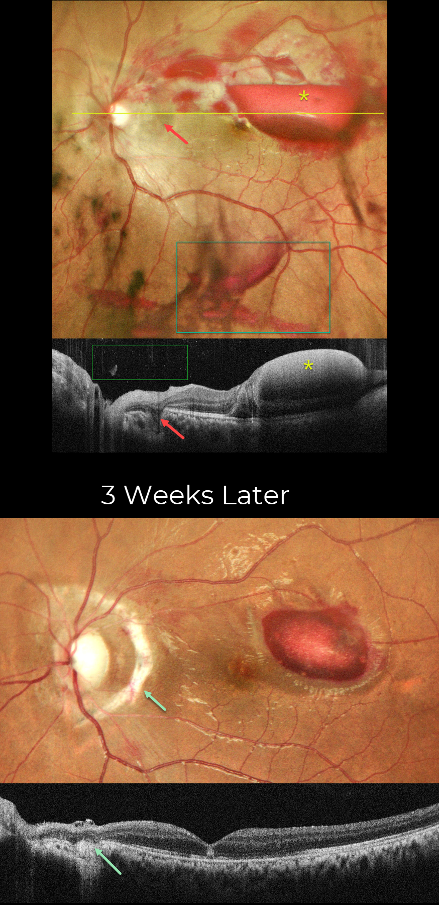
This 19YO male presented with vision loss in his left eye 2 days following blunt trauma from a tennis ball. Vision was counting fingers. Slit lamp examination revealed anterior chamber inflammation, traumatic mydriasis, and nasal subluxation of the crystalline lens.
Pseudocolor SLO imaging shows scattered retinal, sub-internal limiting membrane (ILM), and vitreous blood. The acute choroidal rupture appears as a crescent of increased subretinal pigmentation that arches around the nerve temporally and superiorly. Superior and superotemporal macular commotio retina is faintly visible. On OCT scanning, the vitreous and sub-ILM blood appear hyperreflective. The choroidal rupture appears as a hyporeflective defect extending from the outer retina to the sclera.
Topical steroids, atropine, and a short course of oral steroids were prescribed. Three weeks later, vision improved to 20/200. A prominent depigmented choroidal rupture with bare sclera surrounds the nerve nasally 180 degrees. The vitreous and retinal hemorrhages are virtually completely resolved, and the sub-ILM is decreasing. On OCT, the choroidal rupture shows loss of outer retinal and RPE band detail, with diffuse underlying choroidal hyperreflectivity. There is variable outer retinal band loss in the macula with a central hyperreflective lesion in the outer nuclear layer.
Learning Points:
An acute submacular hemorrhage following blunt trauma almost always indicates an underlying choroidal rupture. As the blood clears, the rupture appears as a curvilinear or crescent-shaped yellowish-white lesion. It occurs due to rapid compression-expansion stress on Bruch’s membrane, which is less elastic and has less tensile strength than the sclera, leading to its fracture. The final vision is usually good unless the rupture extends through the macular center. These patients need to be followed long-term due to the increased risk of macular neovascularization.



