

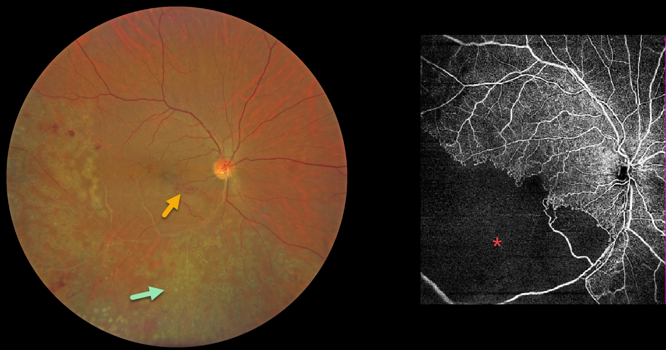
This 42YO male presented with a 3-month history of decreased vision in his right eye. He had received laser treatment in this eye elsewhere. Vision was 20/60 OD.
Color photography shows an old ischemic inferotemporal branch retinal vein occlusion (BRVO) with heavy scatter laser throughout its distribution. Collateral vessels are noted in the inferonasal macula. OCT angiography shows profound capillary loss. There was no macular edema, and observation was recommended.
Learning Points:
In 1986, the Branch Vein Occlusion Study Group reported that scatter laser decreased the risk of vitreous hemorrhage from retinal neovascularization in BRVO and suggested that laser should be applied once neovascularization develops (Arch Ophthalmol 1986;104:34-41). Thirty-five years later, scatter laser remains the gold standard, although some may prefer to stabilize the neovascularization with anti-VEGF therapy before laser.



