

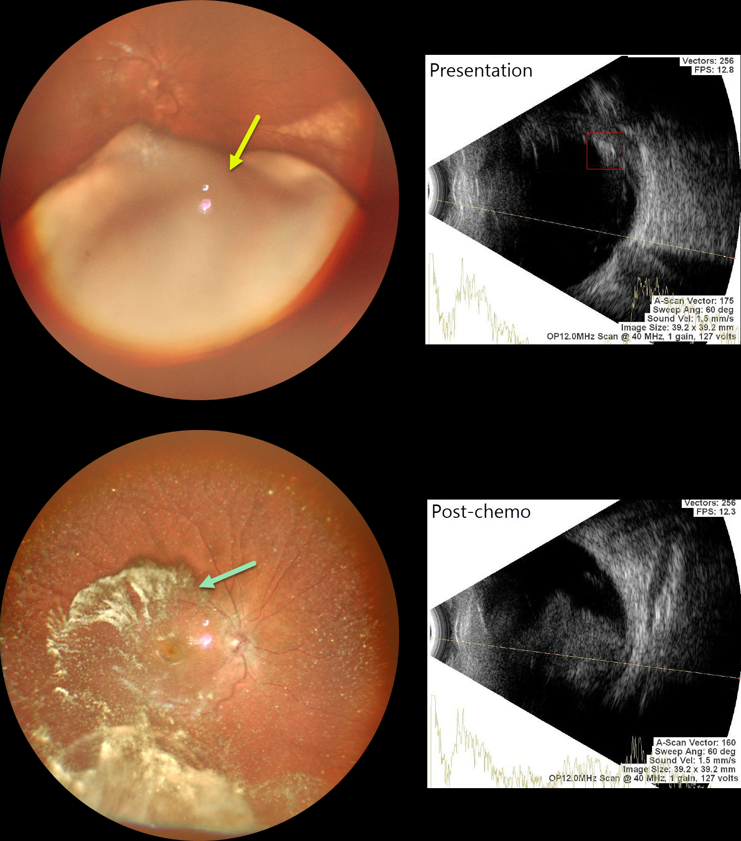
This previously healthy 2YO girl presented with leukocoria in her right eye. Family history was negative.
Examination under anesthesia revealed a large smooth endophytic lesion filling more than half of the vitreous cavity, extending into the inferior macula. B-scan ultrasonography revealed intralesional calcifications. The left eye was normal.
MRI showed no extra-ocular or CNS lesions. We diagnosed her with ICRB Group D retinoblastoma in the right eye. She underwent 3 cycles of chemotherapy.
Repeat examination under anesthesia, 9 weeks following initial presentation, showed marked tumor regression, although there was widespread significant vitreous seeding. MRI did not show any optic nerve infiltration or CNS spread. Enucleation of this eye was scheduled
Learning Points:
Retinoblastoma is the most common primary intraocular malignancy of childhood, with a peak incidence before age five. Presenting signs frequently include leukocoria and strabismus. The disease may be heritable or nonheritable, and delayed diagnosis can result in orbital extension, metastatic spread, and decreased survival. Early identification and intervention with chemotherapy, focal therapies such as laser photocoagulation or cryotherapy, and, in select cases, intra-arterial or intravitreal chemotherapy maximize the chances of ocular salvage and life preservation.



