

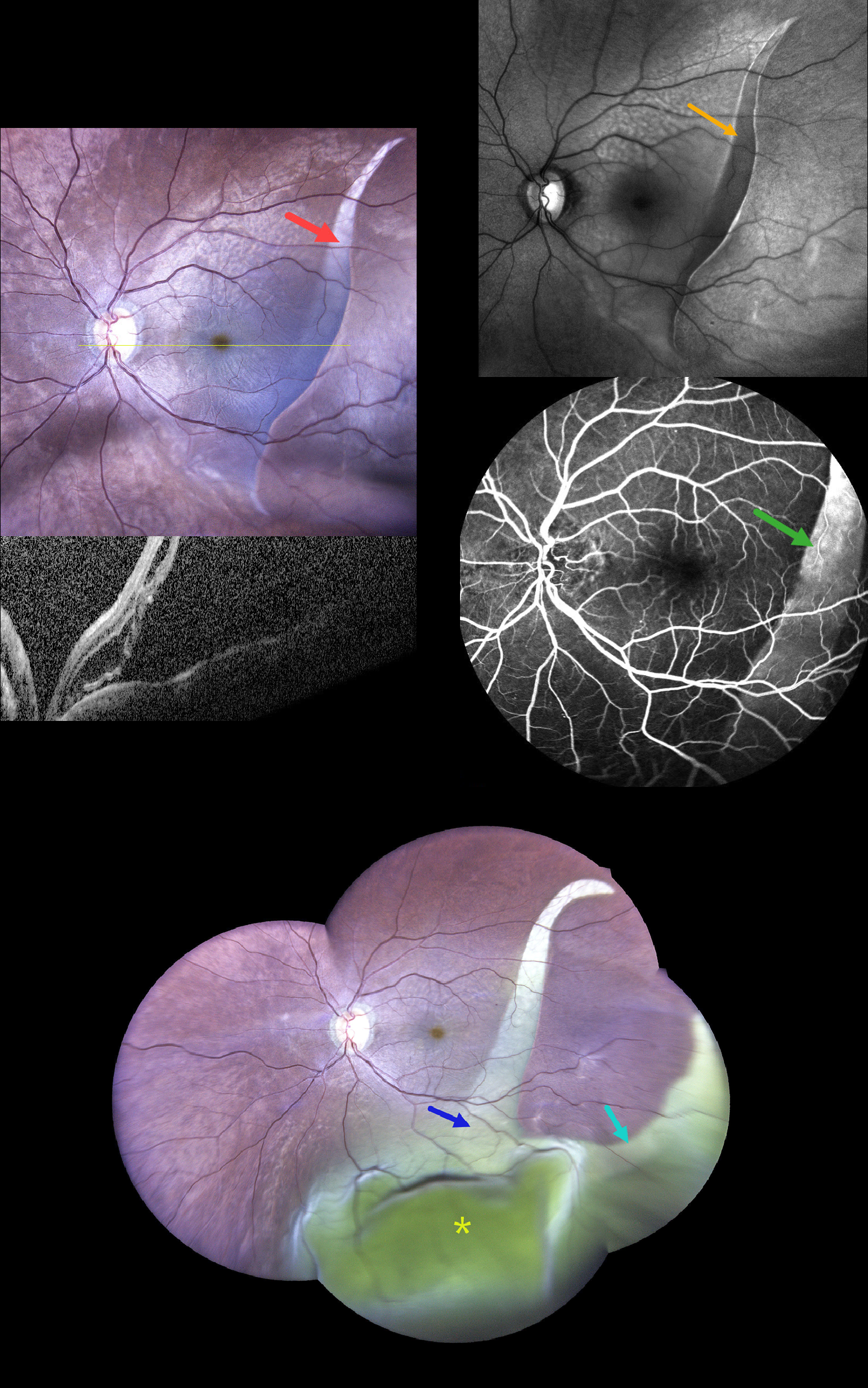
This 68YO male presented with sudden, painless decreased vision in his left eye 2 days ago. He gave a history of type 2 diabetes. Vision was 20/30 (6/9) in his normal OD and hand motion OS.
Widefield photography shows a large curvilinear area of pigment loss (giant RPE tear) extending from the superotemporal into the inferior midperiphery. OCT scanning shows marked macular subretinal fluid. On fundus autofluorescence (FAF), the lesion is hypo-FAF with a corresponding window defect on fluorescein angiography.
He returned 6 days later without new symptoms and stable vision. The prior RPE tear is enlarged in width, extends more inferiorly, and has developed a new branch extending into the inferotemporal midperiphery. A large inferior exudative retinal detachment is noted. Unfortunately, he was immediately lost to follow-up.
Learning Points:
A tear of the RPE is most often seen in RPE detachments associated with neovascular AMD, but can also occur in numerous other etiologies, including idiopathic central serous chorioretinopathy (ICSC) and polypoidal choroidal vasculopathy. The tear leaves a depigmented region where the RPE was and is now absent, along with a pigmented, relatively straight subretinal lesion from the scrolled RPE edge. Our patient’s RPE tear is atypical due to the lack of this hyperpigmented edge, indicating it is more of an RPE dehiscence than a scrolled tear, as well as the multibranched extension that developed 1 week later.
Giant retinal pigment epithelial (RPE) tears occurring outside the macula, defined as tears exceeding 3 clock hours, are most common following trauma or in bullous ICSC and polypoidal choroidal vasculopathy. We have seen these giant tears in previously clinically normal eyes, which later showed pachychoroid-related findings on ICG angiography and en face OCT.



