

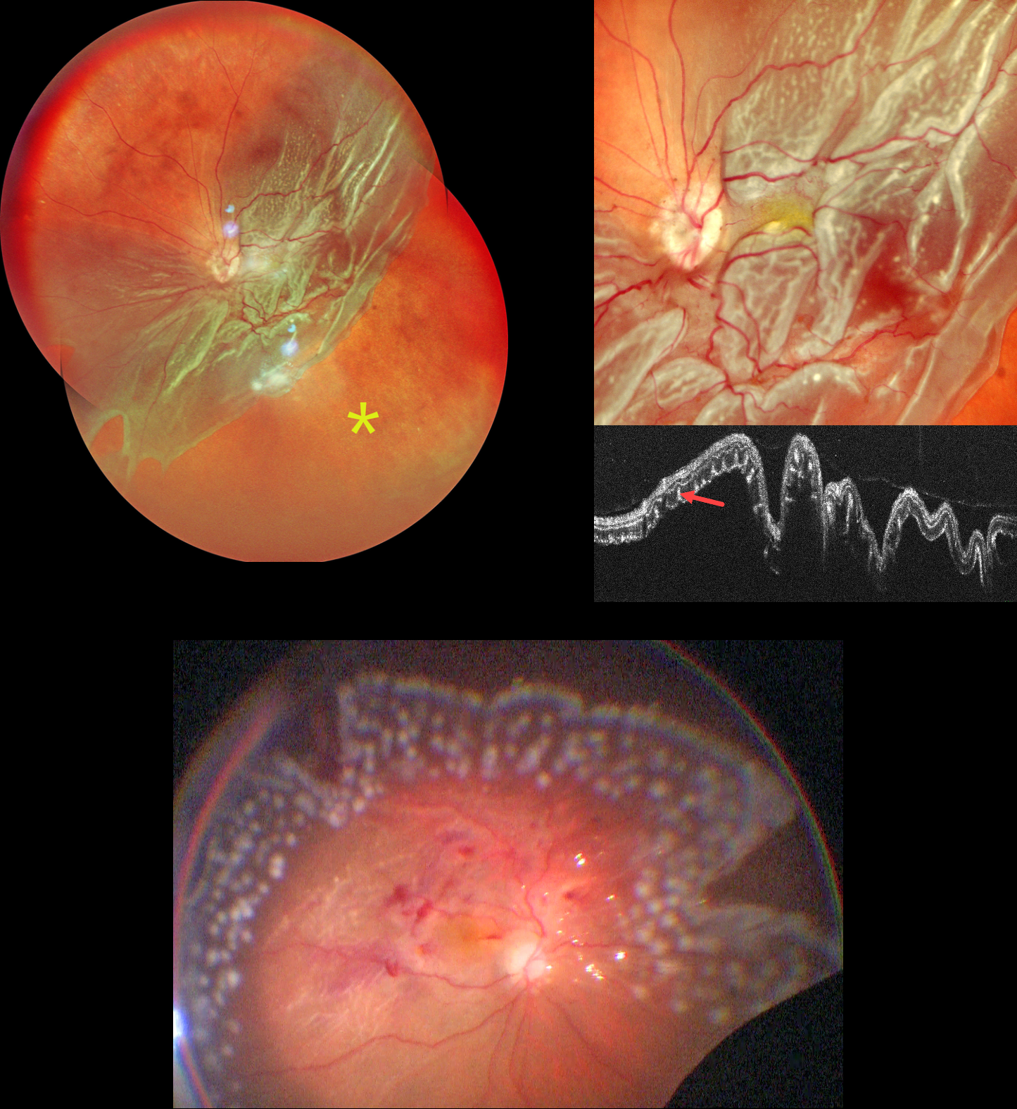
This 62YO male presented with 3 months of vision loss in his left eye. Vision was counting fingers.
Pseudocolor SLO imaging shows a giant retinal tear extending from the 2 to 8 o’clock meridians with bare RPE distal to the posterior retina. There are extensive outer retinal corrugations within the posteriorly detached retina. OCT dramatically shows the detached retina, which is redundantly folded onto itself. The outer retinal corrugations appear as numerous hyperreflective outer retinal pillars.
The detachment was emergently repaired with vitrectomy, membrane peeling, endolaser photocoagulation, and silicone oil. The retina was completely attached at the end of the procedure.
Learning Points:
Outer retinal corrugations, also known as hydration outer retinal folds, are one of the key findings that help differentiate a rhegmatogenous retinal detachment from exudative and traction retinal detachments. The cause of these folds remains uncertain but may result from the hydration of glycosaminoglycans in the interphotoreceptor matrix (Dalvin et al, Retinal Cases & Brief Reports 2020;14:355-359).



