

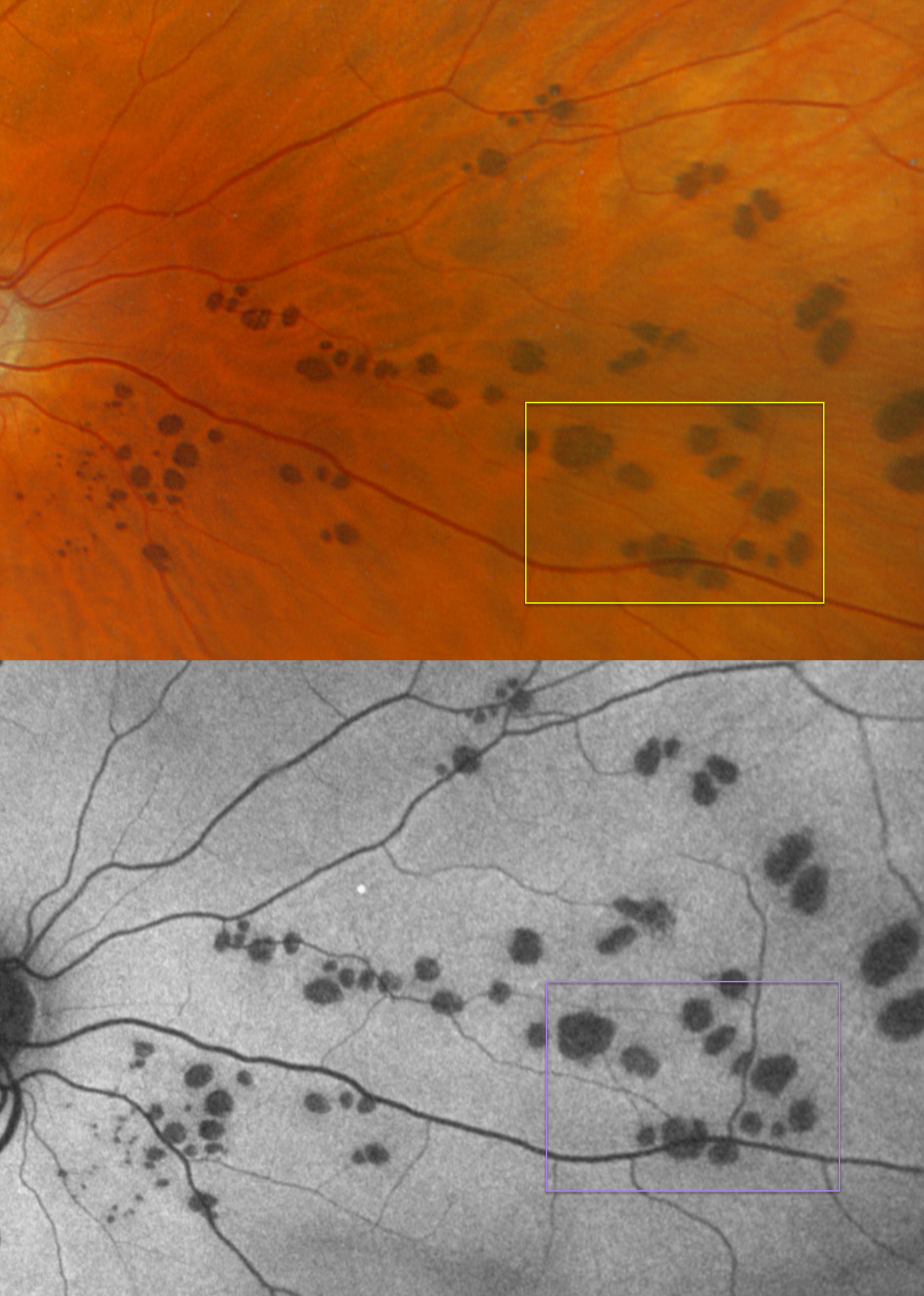
This healthy 59YO male was referred for asymptomatic lesions in his right eye.
Optos color RGB imaging shows multiple variably round-to-oval subretinal pigmented lesions scattered throughout the nasal right fundus. These lesions are hypo-autofluorescent.
Learning Points:
Group-type congenital pigmented nevi of the RPE (bear tracks) is a relatively rare congenital condition characterized by well-demarcated, hyperpigmented, flat, variably sized RPE lesions that resemble bear footprints. Histopathologically, these lesions resemble congenital hypertrophy of the RPE (CHRPE), with increased numbers of pigment granules in normal-sized RPE cells. These lesions are benign and cause no visual symptoms.
The major fluorophore in fundus autofluorescence (FAF) is lipofuscin, which is intraliposomal RPE material generated as byproducts of outer segment metabolism. The outer retina overlying these bear track lesions shows varying degrees of atrophy. This leads to less lipofuscin production with hypo-FAF.
Although bear tracks and the typical CHRPE lesions (large, round, unilateral, with depigmented lacunae with age) are not associated with systemic abnormalities, the multifocal, often bilateral and small comet-shaped lesions with FAP are associated with colonic polyps and cancer (Romania et al, Ophthalmology 1992;99:911-913).



